
Blood in Urine? Understanding the Key Symptom of Bladder Cancer
Bladder cancer stands as one of the most common urological malignancies affecting millions of people worldwide. This comprehensive guide delves into the various aspects of bladder cancer, including its causes, symptoms, diagnostic procedures, and treatment options, providing readers with a thorough understanding of this condition.
Introduction to Bladder Cancer
Bladder cancer originates in the tissues of the bladder, the hollow organ in the lower abdomen that stores urine. The bladder wall consists of several layers, and cancer can begin in any of these layers, though most commonly it starts in the innermost lining, the urothelium. As the cancer progresses, it can grow deeper into the bladder wall and potentially spread to other parts of the body.
According to global cancer statistics, bladder cancer ranks as the tenth most common cancer worldwide, with approximately 573,000 new cases diagnosed annually. It is more prevalent in men than in women, with men being about three to four times more likely to develop this disease. The average age at diagnosis is 73 years, and it is rarely found in people under 40.

Understanding this disease is crucial for early detection and effective treatment. This guide aims to provide comprehensive information about bladder cancer, empowering patients, caregivers, and healthcare professionals with knowledge about this condition.
The Anatomy and Function of the Bladder
To fully grasp bladder cancer, it’s essential to understand the bladder’s structure and function. The bladder is a muscular, expandable organ located in the pelvis, behind the pubic bone and above the prostate in men. In women, it is positioned in front of the uterus and upper vagina.
The bladder’s primary function is to store urine produced by the kidneys before it is eliminated from the body. Urine travels from the kidneys to the bladder through two tubes called ureters. When the bladder is full, nerve signals are sent to the brain, triggering the urge to urinate. During urination, the bladder muscles contract, pushing urine out through the urethra.
The bladder wall consists of four main layers:
1. Urothelium (or transitional epithelium): The innermost layer that directly contacts urine. It is made up of transitional cells that can expand and contract as the bladder fills and empties.
2. Lamina propria: A thin layer of connective tissue beneath the urothelium, containing blood vessels, nerves, and, in some areas, glands.
3. Muscularis propria: The thick layer of muscle tissue that contracts during urination to empty the bladder.
4. Perivesical fat: The outermost layer of fatty tissue that separates the bladder from nearby organs.
Bladder cancer typically begins in the urothelium and can grow deeper into the bladder wall if left untreated. The depth of invasion plays a crucial role in determining the stage of cancer and the appropriate treatment approach.
Types of Bladder Cancer
Bladder cancer is classified based on the type of cells where the cancer originates. The main types include:
A. Urothelial Carcinoma (Transitional Cell Carcinoma)
Urothelial carcinoma is the most common type of bladder cancer, accounting for approximately 90% of all cases. It begins in the urothelial cells that line the inside of the bladder. These cells are called transitional cells because they can change shape without damage when the bladder is full or empty.
Urothelial carcinoma is further divided into two subtypes:
1. Non-invasive urothelial carcinoma: The cancer is confined to the urothelium and has not invaded deeper layers of the bladder wall.
2. Invasive urothelial carcinoma: The cancer has grown from the urothelium into deeper layers of the bladder wall, potentially reaching the muscularis propria or beyond.
B. Squamous Cell Carcinoma
Squamous cell carcinoma accounts for about 4% of bladder cancers in the United States but is more common in regions where schistosomiasis (a parasitic infection) is prevalent. This type of cancer develops in the flat, thin squamous cells that may form in the bladder in response to chronic irritation or inflammation. Most squamous cell carcinomas are invasive.
C. Adenocarcinoma
Adenocarcinoma is a rare type of bladder cancer, making up only about 2% of cases. It begins in glandular cells that release mucus and other fluids. Adenocarcinomas are typically invasive and may be more challenging to treat than urothelial carcinomas.
Other Rare Types
Several other rare types of bladder cancer exist, including:
– Small cell carcinoma: A highly aggressive type that resembles small cell lung cancer.
– Sarcoma: Originates in the bladder’s muscle or fatty tissue.
– Secondary bladder cancer: Cancer that has spread to the bladder from another part of the body.
Understanding the specific type of bladder cancer is crucial for determining the most effective treatment approach, as different types may respond differently to various therapies.
Causes and Risk Factors of Bladder Cancer
While the exact cause of bladder cancer is not always clear, several factors can increase a person’s risk of developing this disease. Understanding these risk factors can help in prevention and early detection.
Tobacco Use
Tobacco use is the single most significant risk factor for bladder cancer, responsible for approximately half of all cases. Smokers are at least three times more likely to develop bladder cancer than non-smokers. The risk increases with the duration and intensity of smoking.
When tobacco smoke is inhaled, harmful chemicals are absorbed into the bloodstream, filtered by the kidneys, and excreted in urine. These carcinogens then damage the cells lining the bladder, potentially leading to cancer over time. Quitting smoking can significantly reduce the risk, though it may take years for the risk to return to that of a non-smoker.
Chemical Exposure
Occupational exposure to certain chemicals, known as aromatic amines, is another significant risk factor for bladder cancer. These chemicals are used in the dye industry and in the production of rubber, leather, textiles, paint products, and certain other materials.
Workers in industries with potential exposure to these carcinogens include:
– Painters
– Mechanics
– Hairdressers
– Truck drivers
– Workers in the rubber, leather, and textile industries
Safety regulations have reduced exposure to these chemicals in many developed countries, but the risk remains for those working without adequate protection. It can take many years after exposure for bladder cancer to develop.
Age and Gender
Bladder cancer risk increases with age, with most cases diagnosed in people over 55. The average age at diagnosis is 73. Men are about three to four times more likely to develop bladder cancer than women, though the reasons for this gender disparity are not fully understood. Some researchers believe that higher rates of smoking and occupational exposure among men may contribute to this difference.
Race and Ethnicity
White people are diagnosed with bladder cancer about twice as often as Black or Hispanic people. Asian Americans have the lowest rates of bladder cancer. These differences may be due to genetic factors, variations in exposure to risk factors, or disparities in healthcare access and utilization.
Chronic Bladder Inflammation
Chronic or repeated urinary tract infections, bladder stones, and other sources of long-term bladder irritation may increase the risk of developing squamous cell carcinoma of the bladder. In many parts of the world, particularly in the Middle East and Africa, chronic infection with the parasite Schistosoma haematobium (schistosomiasis) is a major risk factor for squamous cell carcinoma.
Personal or Family History
Individuals who have previously had bladder cancer are at increased risk of developing it again. Additionally, people with a family history of bladder cancer, especially in a first-degree relative (parent, sibling, or child), may have a higher risk. While most bladder cancers are not inherited, certain genetic syndromes can increase susceptibility.
Previous Cancer Treatment
Treatment with the chemotherapy drug cyclophosphamide (used for various cancers and autoimmune conditions) can increase the risk of bladder cancer. Radiation therapy directed at the pelvis for other cancers may also slightly increase the risk of developing bladder cancer later in life.
Birth Defects of the Bladder
Certain congenital abnormalities of the bladder, such as a persistent urachus (a connection between the umbilicus and bladder that normally disappears before birth), can increase the risk of adenocarcinoma of the bladder.
Fluid Intake
Some studies suggest that drinking plenty of fluids, particularly water, may help lower the risk of bladder cancer. The theory is that increased fluid intake leads to more frequent urination, which may reduce the time that potential carcinogens are in contact with the bladder lining.
Diet
The relationship between diet and bladder cancer risk is not entirely clear, but some research suggests that a diet rich in fruits and vegetables may be protective. Conversely, processed meats and foods high in nitrates or nitrites may increase risk, though more research is needed to confirm these associations.
Other Potential Risk Factors
Other factors that may be associated with an increased risk of bladder cancer include:
– Diabetes
– Certain medications (such as pioglitazone, used to treat diabetes)
– Arsenic in drinking water
– Chlorinated byproducts in drinking water
It’s important to note that having one or more risk factors does not guarantee that a person will develop bladder cancer. Many people with bladder cancer have no known risk factors, while many people with risk factors never develop the disease.
Symptoms of Bladder Cancer
The symptoms of bladder cancer can vary depending on the stage and type of cancer. In its early stages, bladder cancer may not cause noticeable symptoms, which is why regular check-ups are important, especially for those at higher risk.
Hematuria (Blood in Urine)
The most common and often first symptom of bladder cancer is hematuria, or blood in the urine. This occurs in about 80-90% of patients with bladder cancer. Hematuria can be either:
1. Gross hematuria: Blood is visible to the naked eye, causing the urine to appear pink, red, or cola-colored.
2. Microscopic hematuria: Blood is not visible but is detected during a urine test under a microscope.
It’s important to note that hematuria is usually painless and may come and go. Even if it disappears, it should never be ignored, as it can be a sign of bladder cancer or other serious conditions.
Changes in Urination Habits
Bladder cancer can cause changes in urination patterns, including:
1. Increased frequency: Needing to urinate more often than usual.
2. Urgency: A sudden, strong urge to urinate that may be difficult to control.
3. Dysuria: Pain or burning during urination.
4. Nocturia: Waking up multiple times during the night to urinate.
5. Hesitancy: Difficulty starting urination.
6. Weak urine stream.
These symptoms are more common in advanced bladder cancer and can be similar to those caused by urinary tract infections, benign prostatic hyperplasia (BPH), or other bladder conditions.
Pelvic or Back Pain
As bladder cancer progresses, it may cause pain in the pelvic area or lower back. This pain can be persistent or intermittent and may range from mild to severe. In advanced cases, the pain may indicate that the cancer has spread to nearby tissues or bones.
Unintended Weight Loss and Fatigue
In advanced stages of bladder cancer, patients may experience unexplained weight loss and persistent fatigue. These symptoms are often associated with many types of cancer and result from the body’s response to the disease.
Other Symptoms
Other less common symptoms of bladder cancer include:
1. Swelling in the feet: This can occur if the cancer blocks the tubes that connect the kidneys to the bladder (ureters), causing urine to back up into the kidneys.
2. Bone pain: This may indicate that the cancer has spread to the bones.
3. Anemia: A low red blood cell count, which can cause fatigue, weakness, and shortness of breath.
It’s crucial to remember that these symptoms can be caused by conditions other than bladder cancer. However, if you experience any of these symptoms, especially blood in your urine, it’s important to consult a healthcare provider for proper evaluation and diagnosis.
Diagnosis of Bladder Cancer
The diagnosis of bladder cancer involves a series of tests and procedures to confirm the presence of cancer, determine its type, and assess its stage. Early diagnosis is crucial for effective treatment and better outcomes.
Initial Evaluation
When a patient presents with symptoms suggestive of bladder cancer, the healthcare provider will begin with:
1. Medical history: The doctor will ask about symptoms, risk factors (such as smoking history or occupational exposures), and any personal or family history of cancer.
2. Physical examination: This may include a digital rectal exam for men or a pelvic exam for women to check for any abnormalities.
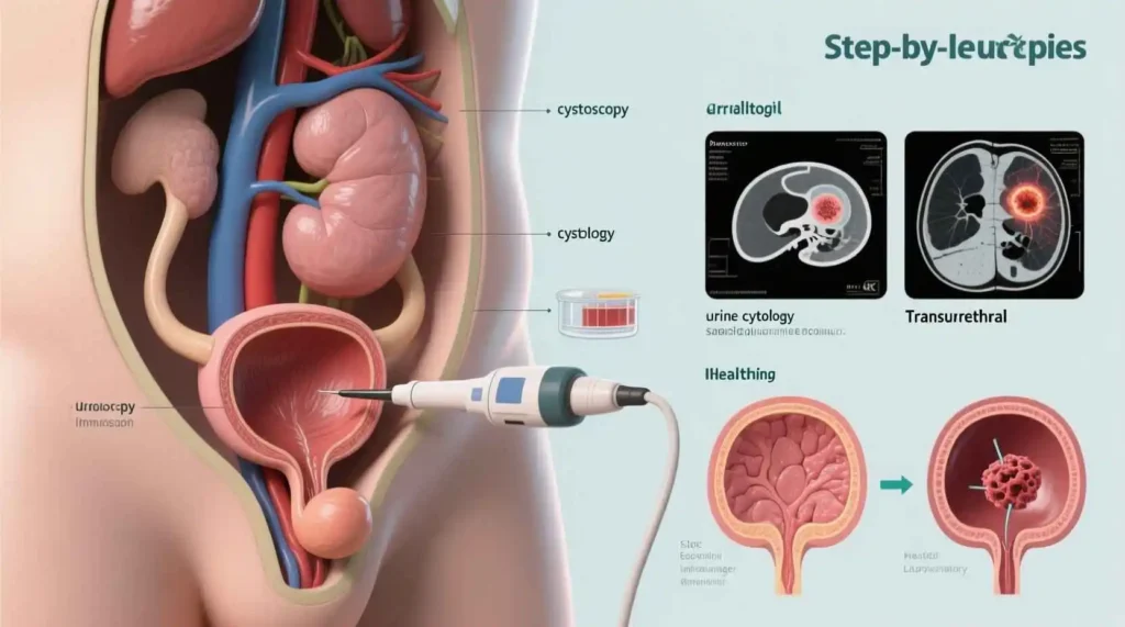
Urine Tests
Several urine tests can help detect bladder cancer or rule out other conditions:
1. Urinalysis: A basic test to check for blood, infection, or other abnormalities in the urine.
2. Urine cytology: A laboratory test in which a urine sample is examined under a microscope for cancer cells. This test is more effective at detecting high-grade bladder cancers but may miss low-grade tumors.
3. Urine biomarker tests: These tests look for specific substances released by bladder cancer cells into the urine. Examples include:
– NMP22 (Nuclear Matrix Protein 22)
– BTA (Bladder Tumor Antigen) tests
– ImmunoCyt
– UroVysion (a fluorescence in situ hybridization test that detects chromosomal abnormalities)
While these tests can be helpful, they are not definitive for diagnosing bladder cancer and are often used in conjunction with other diagnostic procedures.
Imaging Tests
Imaging tests provide detailed pictures of the urinary tract and can help identify tumors and determine if cancer has spread:
1. Ultrasound: This non-invasive test uses sound waves to create images of the bladder and surrounding structures. It can detect tumors and blockages in the urinary tract.
2. Computed Tomography (CT) scan: A CT urogram uses contrast dye to create detailed cross-sectional images of the urinary tract, including the kidneys, ureters, and bladder. This test can identify tumors and assess if cancer has spread to other areas.
3. Magnetic Resonance Imaging (MRI): MRI uses powerful magnets and radio waves to create detailed images of the bladder and surrounding tissues. It is particularly useful for determining the depth of tumor invasion into the bladder wall.
4. Intravenous Pyelogram (IVP): This older imaging technique involves injecting contrast dye into a vein, which then travels to the urinary tract and makes the kidneys, ureters, and bladder visible on X-rays. It has largely been replaced by CT urography.
5. Chest X-ray or CT scan: These tests may be performed to check if bladder cancer has spread to the lungs.
Cystoscopy
Cystoscopy is the gold standard for diagnosing bladder cancer. During this procedure:
1. The doctor inserts a thin, flexible tube with a light and camera (cystoscope) through the urethra into the bladder.
2. The bladder is filled with a sterile solution to expand it and allow for better visualization.
3. The doctor examines the bladder lining for any abnormalities, tumors, or suspicious areas.
4. If abnormalities are found, the doctor may take a small tissue sample (biopsy) or remove the entire tumor during the procedure.
Cystoscopy can be performed in a doctor’s office under local anesthesia or in an operating room under general or spinal anesthesia, depending on the complexity of the procedure.
Biopsy and Pathology
If a suspicious area is found during cystoscopy, a biopsy is performed to confirm the diagnosis of bladder cancer. The tissue sample is sent to a pathologist, who examines it under a microscope to determine:
1. Whether cancer is present.
2. The type of cancer (urothelial carcinoma, squamous cell carcinoma, adenocarcinoma, etc.).
3. The grade of the cancer (how abnormal the cells look under a microscope):
– Low-grade: The cells look more like normal bladder tissue and tend to grow slowly.
– High-grade: The cells look very abnormal and are more likely to grow quickly and spread.
4. The depth of invasion (how far the cancer has grown into the bladder wall).
Transurethral Resection of Bladder Tumor (TURBT)
For patients with suspected bladder cancer, a TURBT is often both a diagnostic and therapeutic procedure. During TURBT:
1. The patient is given general or spinal anesthesia.
2. A special type of cystoscope called a resectoscope is inserted into the bladder.
3. The surgeon removes the tumor and a small margin of surrounding healthy tissue.
4. The tissue is sent to a pathologist for examination.
5. In some cases, the surgeon may also perform fulguration, which uses an electric current to burn away any remaining cancer cells.
TURBT allows for accurate staging and grading of the cancer and is often the first step in treatment for non-muscle invasive bladder cancer.
Staging of Bladder Cancer
Once bladder cancer is diagnosed, it is staged to determine the extent of the disease. Staging is crucial for determining the most appropriate treatment approach. The most commonly used staging system is the TNM system, which stands for:
– T (Tumor): The size and extent of the primary tumor.
– N (Node): Whether cancer has spread to nearby lymph nodes.
– M (Metastasis): Whether cancer has spread to distant parts of the body.
T Stages (Tumor)
– Ta: Non-invasive papillary carcinoma (the tumor is confined to the urothelium).
– Tis: Carcinoma in situ (flat, non-invasive cancer confined to the urothelium).
– T1: The tumor has grown into the connective tissue beneath the urothelium (lamina propria) but not into the muscle layer.
– T2: The tumor has grown into the muscularis propria (muscle layer).
– T2a: The tumor has grown into the inner half of the muscle layer.
– T2b: The tumor has grown into the outer half of the muscle layer.
– T3: The tumor has grown through the muscle layer into the fatty tissue surrounding the bladder.
– T3a: The tumor is microscopic.
– T3b: The tumor is visible to the naked eye (macroscopic).
– T4: The tumor has grown into nearby organs or structures.
– T4a: The tumor has grown into the prostate, seminal vesicles, uterus, or vagina.
– T4b: The tumor has grown into the pelvic wall or abdominal wall.
N Stages (Node)
– N0: No cancer in nearby lymph nodes.
– N1: Cancer has spread to a single lymph node in the pelvis.
– N2: Cancer has spread to two or more lymph nodes in the pelvis.
– N3: Cancer has spread to lymph nodes along the common iliac artery.
M Stages (Metastasis)
– M0: No distant metastasis.
– M1: Distant metastasis is present.
– M1a: Cancer has spread to distant lymph nodes beyond the pelvis.
– M1b: Cancer has spread to other distant organs, such as the lungs, bones, or liver.
Overall Stages
Based on the TNM classification, bladder cancer is assigned an overall stage:
– Stage 0a: Ta, N0, M0 (non-invasive papillary carcinoma)
– Stage 0is: Tis, N0, M0 (carcinoma in situ)
– Stage I: T1, N0, M0
– Stage II: T2a or T2b, N0, M0
– Stage III: T3a, T3b, or T4a, N0, M0; or any T, N1 or N2, M0
– Stage IV: T4b, any N, M0; or any T, N3, M0; or any T, any N, M1
The stage of bladder cancer is a critical factor in determining the prognosis and selecting the most appropriate treatment approach.
Treatment of Bladder Cancer
The treatment of bladder cancer depends on several factors, including the stage and grade of the cancer, the patient’s overall health, and personal preferences. Treatment options range from minimally invasive procedures for early-stage cancer to more extensive surgeries and systemic therapies for advanced disease.
Non-Muscle Invasive Bladder Cancer (NMIBC) Treatment
Non-muscle invasive bladder cancer (stages Ta, T1, and Tis) is confined to the inner layers of the bladder wall and has not invaded the muscle layer. The primary treatment for NMIBC is transurethral resection of bladder tumor (TURBT), often followed by intravesical therapy to reduce the risk of recurrence.
Transurethral Resection of Bladder Tumor (TURBT)
TURBT is both a diagnostic and therapeutic procedure for NMIBC. During TURBT, the surgeon removes the tumor and a small margin of surrounding healthy tissue. The procedure is performed through the urethra, without the need for external incisions.
After TURBT, the pathologist examines the removed tissue to determine the stage and grade of the cancer. This information guides further treatment decisions.
Intravesical Therapy
Intravesical therapy involves placing liquid medication directly into the bladder through a catheter. This allows the medication to come into direct contact with the bladder lining while minimizing effects on the rest of the body. Intravesical therapy is typically started a few weeks after TURBT and may be given as a single dose or as a series of treatments over several weeks or months.
The two main types of intravesical therapy are: