

Understanding Blood: The Lifesaving Fluid and Its Critical Components
Human blood is one of the most remarkable substances in the human body, a living tissue that performs countless essential functions to keep us alive and healthy. This complex fluid circulates through our vascular system, delivering oxygen and nutrients to cells, removing waste products, fighting infections, and maintaining homeostasis. In this comprehensive exploration of human blood, we will delve into its composition, functions, production, and the vital role it plays in human health and disease.
Introduction to Human Blood
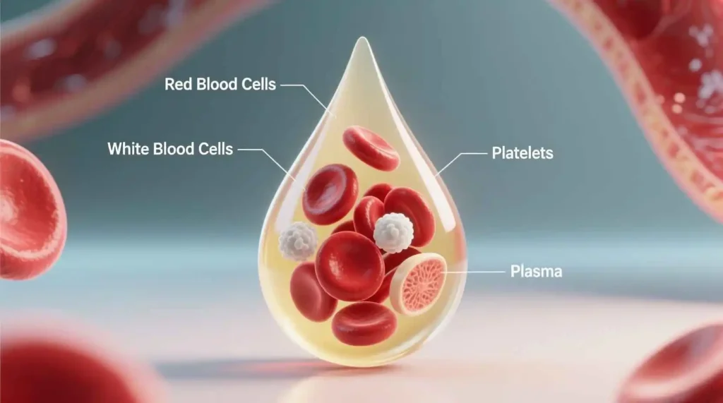
Blood is a specialized bodily fluid that delivers necessary substances such as nutrients and oxygen to the cells and transports metabolic waste products away from those same cells. In vertebrates, it is composed of blood cells suspended in blood plasma. Plasma, which constitutes 55% of blood fluid, is mostly water (92% by volume), and contains proteins, glucose, mineral ions, hormones, carbon dioxide, and blood cells themselves.
The average adult has a blood volume of approximately 5 liters, which accounts for about 7-8% of total body weight. Blood circulates through the cardiovascular system, propelled by the pumping action of the heart, and plays a critical role in maintaining the body’s overall health and functioning.

Blood has been recognized as a vital fluid since ancient times, with early civilizations associating it with life itself. The Roman physician Galen believed that blood was produced in the liver and distributed throughout the body, a theory that persisted until the 17th century when William Harvey described the circulation of blood. Today, our understanding of blood has advanced tremendously, allowing us to appreciate its complexity and importance.
The Composition of Blood
Blood is a complex mixture of cellular components suspended in a liquid matrix. To understand blood fully, we must examine both its cellular components and the plasma in which they float.
### Blood Plasma
Plasma is the liquid component of blood, a straw-colored fluid that constitutes about 55% of total blood volume. It is primarily composed of water (about 92%), with the remaining 8% consisting of various dissolved substances including:
1. Proteins: Plasma proteins make up about 7% of plasma and serve numerous functions. The major plasma proteins include:
– Albumin: This is the most abundant plasma protein, produced by the liver. Albumin helps maintain osmotic pressure, which prevents fluid from leaking out of blood vessels into tissues. It also serves as a carrier protein for various substances, including hormones, fatty acids, and drugs.
– Globulins: These proteins are divided into alpha, beta, and gamma globulins. Alpha and beta globulins are produced by the liver and function as transport proteins for lipids, hormones, and minerals. Gamma globulins are antibodies produced by plasma cells and are essential for immune function.
– Fibrinogen: This protein is produced by the liver and plays a crucial role in blood clotting. When blood vessels are damaged, fibrinogen is converted to fibrin, which forms a mesh that traps blood cells to form a clot.
2. Electrolytes: Plasma contains various electrolytes, including sodium, potassium, calcium, magnesium, chloride, and bicarbonate. These ions are essential for maintaining fluid balance, pH regulation, nerve conduction, and muscle contraction.
3. Nutrients: Plasma transports nutrients absorbed from the digestive system to cells throughout the body. These include glucose, amino acids, fatty acids, glycerol, vitamins, and minerals.
4. Waste Products: Metabolic waste products are carried in the blood to organs of excretion. These include urea and creatinine (excreted by the kidneys), bilirubin (excreted by the liver), and carbon dioxide (excreted by the lungs).
5. Hormones: Endocrine glands secrete hormones directly into the blood, which transports them to target organs where they exert their effects.
6. Gases: Plasma contains dissolved gases, primarily oxygen and carbon dioxide, which are transported to and from tissues.
Cellular Components of Blood
The cellular components of blood, collectively called formed elements, make up about 45% of blood volume. These include red blood cells, white blood cells, and platelets. The percentage of blood volume occupied by red blood cells is called the hematocrit, which is normally about 45% for men and 40% for women.
#### Red Blood Cells (Erythrocytes)
Red blood cells, or erythrocytes, are the most abundant cells in blood, accounting for about 99% of all blood cells. A microliter of blood contains approximately 4.5-5.5 million red blood cells in men and 4.0-5.0 million in women.
Red blood cells are unique in several ways:
1. Structure: Mature red blood cells are biconcave discs, a shape that increases their surface area for gas exchange and allows them to flex and squeeze through narrow capillaries. They lack a nucleus and most organelles, which provides more space for hemoglobin, the oxygen-carrying protein.
2. Hemoglobin: Each red blood cell contains about 280 million hemoglobin molecules. Hemoglobin is a complex protein composed of four polypeptide chains, each with an iron-containing heme group that can bind one oxygen molecule. This structure allows each hemoglobin molecule to carry four oxygen molecules.
3. Function: The primary function of red blood cells is to transport oxygen from the lungs to tissues and carbon dioxide from tissues to the lungs. Oxygen binds to hemoglobin in the lungs, where oxygen concentration is high, and is released in tissues, where oxygen concentration is low. Carbon dioxide is transported in three ways: dissolved in plasma, bound to hemoglobin, and as bicarbonate ions.
4. Production: Red blood cells are produced in the bone marrow through a process called erythropoiesis. The process begins with hematopoietic stem cells, which differentiate into erythroblasts, then reticulocytes, and finally mature erythrocytes. This process is regulated by erythropoietin, a hormone produced primarily by the kidneys in response to hypoxia (low oxygen levels).
5. Lifespan and Destruction: Red blood cells have a lifespan of about 120 days. As they age, they become less flexible and are removed from circulation by macrophages in the spleen and liver. The iron from hemoglobin is recycled, while the heme portion is converted to bilirubin, which is excreted in bile.
#### White Blood Cells (Leukocytes)
White blood cells, or leukocytes, are the cellular components of blood that protect the body against infectious diseases and foreign invaders. Unlike red blood cells, white blood cells have a nucleus and other organelles and can move independently in and out of blood vessels.
White blood cells are much less numerous than red blood cells, with a normal range of 4,000-11,000 cells per microliter of blood. They are classified into two main groups based on the presence or absence of granules in their cytoplasm: granulocytes and agranulocytes.
1. Granulocytes:
– Neutrophils: These are the most abundant white blood cells, accounting for 50-70% of all leukocytes. They have a multi-lobed nucleus and fine granules that stain pale lilac. Neutrophils are the first responders to bacterial infections, where they engulf and destroy pathogens through phagocytosis. They have a short lifespan of only a few hours to a few days.
– Eosinophils: These cells make up 1-4% of white blood cells and have a bilobed nucleus and large granules that stain red with eosin dye. Eosinophils play a role in combating parasitic infections and are involved in allergic reactions and asthma.
– Basophils: The rarest of the white blood cells (less than 1%), basophils have a bilobed nucleus and large granules that stain blue with basic dyes. They release histamine and other mediators of inflammation during allergic reactions and play a role in protecting against parasites.
2. Agranulocytes:
– Lymphocytes: These cells account for 20-40% of white blood cells and have a large, round nucleus that occupies most of the cell. Lymphocytes are key components of the immune system and are divided into three main types:
– B cells: These cells produce antibodies that bind to specific antigens on pathogens, marking them for destruction.
– T cells: These cells coordinate the immune response and directly kill infected or cancerous cells.
– Natural killer (NK) cells: These cells provide rapid responses to virus-infected cells and tumor cells.
– Monocytes: These are the largest white blood cells, accounting for 2-8% of leukocytes. They have a kidney-shaped nucleus and abundant cytoplasm. Monocytes circulate in the blood for about 1-3 days before migrating into tissues, where they differentiate into macrophages or dendritic cells. Macrophages are phagocytic cells that engulf pathogens, dead cells, and cellular debris, while dendritic cells present antigens to T cells, initiating an immune response.
#### Platelets (Thrombocytes)
Platelets, or thrombocytes, are not actually cells but small, disc-shaped cell fragments that play a crucial role in blood clotting. They are produced in the bone marrow from large cells called megakaryocytes.
1. Structure: Platelets are much smaller than red or white blood cells, measuring only 2-3 micrometers in diameter. They have no nucleus but contain granules with various clotting factors and enzymes.
2. Function: When a blood vessel is damaged, platelets adhere to the site of injury, become activated, and aggregate to form a temporary plug. They also release substances that promote blood clotting and vessel repair.
3. Production: Platelets are produced in the bone marrow through a process called thrombopoiesis. Hematopoietic stem cells differentiate into megakaryoblasts, which mature into megakaryocytes. Megakaryocytes extend long processes called proplatelets into blood vessels, where platelets break off and enter circulation.
4. Lifespan: Platelets have a lifespan of about 7-10 days. Aged or damaged platelets are removed from circulation by macrophages in the spleen and liver.
The Functions of Blood
Blood performs numerous essential functions that are vital for maintaining health and homeostasis. These functions can be broadly categorized into transportation, regulation, and protection.
### Transportation Functions
1. Oxygen Transport: Red blood cells transport oxygen from the lungs to all body tissues. Oxygen binds to hemoglobin in the lungs, where oxygen concentration is high, and is released in tissues, where oxygen concentration is low. This process is facilitated by the cooperative binding of oxygen to hemoglobin, which allows for efficient loading and unloading of oxygen.
2. Carbon Dioxide Transport: Blood transports carbon dioxide, a waste product of cellular metabolism, from tissues to the lungs for elimination. Carbon dioxide is transported in three ways:
– Dissolved in plasma (about 7%)
– Bound to hemoglobin as carbaminohemoglobin (about 23%)
– As bicarbonate ions (about 70%), formed when carbon dioxide reacts with water in red blood cells, a reaction catalyzed by the enzyme carbonic anhydrase
3. Nutrient Transport: Blood carries nutrients absorbed from the digestive system to cells throughout the body. These include glucose, amino acids, fatty acids, glycerol, vitamins, and minerals. These nutrients are either dissolved in plasma or bound to carrier proteins.
4. Hormone Transport: Endocrine glands secrete hormones directly into the blood, which transports them to target organs where they exert their effects. For example, insulin secreted by the pancreas is transported by blood to cells throughout the body, where it promotes glucose uptake.
5. Waste Product Transport: Blood carries metabolic waste products to organs of excretion. Urea and creatinine are transported to the kidneys for excretion in urine, bilirubin is transported to the liver for excretion in bile, and carbon dioxide is transported to the lungs for exhalation.
### Regulation Functions
1. Temperature Regulation: Blood helps regulate body temperature by distributing heat throughout the body. When body temperature rises, blood vessels in the skin dilate, increasing blood flow to the skin and promoting heat loss through radiation and convection. When body temperature falls, blood vessels in the skin constrict, reducing blood flow to the skin and conserving heat.
2. pH Regulation: Blood helps maintain the body’s acid-base balance through various buffering systems. The most important of these is the bicarbonate buffer system, which consists of carbonic acid and bicarbonate ions. When blood pH becomes too acidic, bicarbonate ions bind with excess hydrogen ions, and when pH becomes too alkaline, carbonic acid releases hydrogen ions. Blood also contains proteins and phosphate buffers that help maintain pH within the narrow range necessary for normal cellular function.
3. Fluid Balance: Blood helps regulate fluid balance between blood vessels and tissues. This is primarily achieved through the osmotic pressure exerted by plasma proteins, particularly albumin, which prevents excessive fluid from leaking out of blood vessels into tissues.
### Protection Functions
1. Immune Defense: White blood cells protect the body against infectious diseases and foreign invaders through various mechanisms:
– Phagocytosis: Neutrophils and monocytes/macrophages engulf and destroy pathogens, dead cells, and cellular debris.
– Antibody Production: B cells produce antibodies that bind to specific antigens on pathogens, marking them for destruction.
– Cell-Mediated Immunity: T cells coordinate the immune response and directly kill infected or cancerous cells.
– Natural Killer Activity: NK cells provide rapid responses to virus-infected cells and tumor cells.
2. Hemostasis: Blood prevents excessive blood loss through a process called hemostasis, which involves three main steps:
– Vascular Spasm: When a blood vessel is damaged, it constricts to reduce blood flow.
– Platelet Plug Formation: Platelets adhere to the site of injury, become activated, and aggregate to form a temporary plug.
– Coagulation: A complex cascade of reactions involving various clotting factors leads to the formation of fibrin, which forms a mesh that traps blood cells to form a stable clot.
3. Prevention of Infection: Blood contains various proteins that help prevent infection, including complement proteins (which enhance phagocytosis and can directly kill pathogens), interferons (which interfere with viral replication), and acute-phase proteins (which are produced in response to inflammation and infection).
Blood Production and Regulation
The production of blood cells, called hematopoiesis, is a complex process that occurs primarily in the bone marrow. This process is tightly regulated to maintain adequate numbers of each type of blood cell.
### Hematopoiesis
Hematopoiesis begins with hematopoietic stem cells (HSCs), which are multipotent stem cells capable of giving rise to all blood cell lineages. These stem cells reside in the bone marrow and can self-renew or differentiate into various progenitor cells.
The process of hematopoiesis can be divided into two main pathways:
1. Myeloid Lineage: This pathway gives rise to red blood cells, platelets, and most white blood cells (neutrophils, eosinophils, basophils, and monocytes). The process begins with myeloid stem cells, which differentiate into various progenitor cells, then into precursor cells, and finally into mature blood cells.
2. Lymphoid Lineage: This pathway gives rise to lymphocytes (B cells, T cells, and NK cells). The process begins with lymphoid stem cells, which differentiate into lymphoid progenitor cells, then into precursor cells, and finally into mature lymphocytes.
During fetal development, hematopoiesis occurs in several locations, including the yolk sac, liver, spleen, and bone marrow. After birth, hematopoiesis is primarily confined to the bone marrow, although the liver and spleen can resume this function in certain disease states.
Regulation of Blood Cell Production
The production of blood cells is regulated by various growth factors and cytokines that stimulate the proliferation and differentiation of hematopoietic stem cells and progenitor cells. These regulatory substances are produced by various cells, including stromal cells in the bone marrow, lymphocytes, macrophages, and endothelial cells.
Key regulators of hematopoiesis include:
1. Erythropoietin (EPO): This hormone is produced primarily by the kidneys in response to hypoxia (low oxygen levels). EPO stimulates the production of red blood cells by promoting the proliferation and differentiation of erythroid progenitor cells. In conditions of chronic hypoxia, such as living at high altitudes or chronic lung disease, EPO production increases, leading to an increase in red blood cell production.
2. Thrombopoietin (TPO): This hormone is produced primarily by the liver and kidneys. TPO stimulates the production of platelets by promoting the proliferation and differentiation of megakaryocytes. In conditions of thrombocytopenia (low platelet count), TPO production increases, leading to an increase in platelet production.
3. Colony-Stimulating Factors (CSFs): These are glycoproteins that stimulate the production and function of white blood cells. Key CSFs include:
– Granulocyte colony-stimulating factor (G-CSF): Stimulates the production of neutrophils.
– Granulocyte-macrophage colony-stimulating factor (GM-CSF): Stimulates the production of granulocytes and monocytes.
– Macrophage colony-stimulating factor (M-CSF): Stimulates the production of monocytes and macrophages.
4. Interleukins (ILs): These are cytokines that play important roles in immune responses and hematopoiesis. Key interleukins involved in hematopoiesis include IL-3 (which stimulates the production of multiple blood cell lineages), IL-5 (which stimulates the production of eosinophils), and IL-7 (which stimulates the production of lymphocytes).
5. Stem Cell Factor (SCF): This cytokine is produced by stromal cells in the bone marrow and plays a crucial role in the maintenance and proliferation of hematopoietic stem cells.
The production of blood cells is also regulated by feedback mechanisms. For example, when red blood cell count increases, oxygen delivery to tissues improves, reducing EPO production and subsequently decreasing red blood cell production. Similarly, when white blood cell count increases after an infection, the production of CSFs decreases, reducing white blood cell production.
Blood Types and Compatibility
Blood types are classifications of blood based on the presence or absence of specific antigens on the surface of red blood cells. The most important blood group systems for blood transfusions are the ABO system and the Rh system.
### The ABO Blood Group System
The ABO blood group system is the most significant blood group system in human blood transfusion. It is determined by the presence or absence of A and B antigens on the surface of red blood cells and the corresponding antibodies in the plasma.
1. Type A: Red blood cells have A antigens on their surface, and the plasma contains anti-B antibodies.
2. Type B: Red blood cells have B antigens on their surface, and the plasma contains anti-A antibodies.
3. Type AB: Red blood cells have both A and B antigens on their surface, and the plasma contains neither anti-A nor anti-B antibodies.
4. Type O: Red blood cells have neither A nor B antigens on their surface, and the plasma contains both anti-A and anti-B antibodies.
The ABO blood group is determined by the ABO gene, which has three alleles: A, B, and O. The A and B alleles are codominant, while the O allele is recessive. This means that:
– Individuals with genotype AA or AO have type A blood.
– Individuals with genotype BB or BO have type B blood.
– Individuals with genotype AB have type AB blood.
– Individuals with genotype OO have type O blood.
The Rh Blood Group System
The Rh blood group system is the second most important blood group system after ABO. It is determined by the presence or absence of the RhD antigen on the surface of red blood cells.
1. Rh-positive (Rh+): Red blood cells have the RhD antigen on their surface.
2. Rh-negative (Rh-): Red blood cells lack the RhD antigen on their surface.
The Rh blood group is determined by the RHD gene. Individuals with at least one copy of the RHD gene are Rh-positive, while those without any copies are Rh-negative.
Blood Type Compatibility
Understanding blood type compatibility is crucial for safe blood transfusions. When a person receives blood that contains antigens their immune system recognizes as foreign, their antibodies will bind to those antigens, causing a transfusion reaction that can be life-threatening.
1. ABO Compatibility:
– Type A individuals can receive blood from type A or type O donors.
– Type B individuals can receive blood from type B or type O donors.
– Type AB individuals can receive blood from type A, type B, type AB, or type O donors (they are universal recipients).
– Type O individuals can receive blood only from type O donors (they are universal donors).
2. Rh Compatibility:
– Rh-positive individuals can receive blood from Rh-positive or Rh-negative donors.
– Rh-negative individuals should receive blood from Rh-negative donors to avoid developing anti-Rh antibodies.
When considering both ABO and Rh compatibility, the universal donor is O-negative (can be given to anyone), and the universal recipient is AB-positive (can receive from anyone).
Other Blood Group Systems
In addition to ABO and Rh, there are many other blood group systems, including Kell, Duffy, Kidd, and MNS. While these systems are less likely to cause transfusion reactions, they can still be important in certain situations, particularly for patients who require multiple transfusions or have developed antibodies against specific antigens.
Blood Disorders
Blood disorders are conditions that affect one or more components of blood. These disorders can be broadly categorized into disorders of red blood cells, white blood cells, platelets, and plasma.
### Red Blood Cell Disorders
1. Anemia: Anemia is a condition characterized by a decrease in the number of red blood cells or the amount of hemoglobin in the blood. There are many types of anemia, including:
– Iron-deficiency anemia: The most common type of anemia, caused by insufficient iron intake, absorption, or utilization.
– Vitamin B12 deficiency anemia: Caused by insufficient intake or absorption of vitamin B12, which is necessary for red blood cell production.
– Folate deficiency anemia: Caused by insufficient intake or absorption of folate, which is necessary for DNA synthesis and red blood cell production.
– Aplastic anemia: A rare condition in which the bone marrow fails to produce enough blood cells.
– Hemolytic anemia: Caused by the premature destruction of red blood cells.
– Sickle cell anemia: An inherited disorder in which red blood cells assume a sickle shape, leading to blockages in blood vessels and reduced oxygen delivery.
– Thalassemia: An inherited disorder in which the body produces an abnormal form of hemoglobin, leading to the destruction of red blood cells.
2. Polycythemia: Polycythemia is a condition characterized by an increase in the number of red blood cells. This can be primary (polycythemia vera, a bone marrow disorder) or secondary (caused by factors such as chronic hypoxia or certain tumors).
3. Malaria: A parasitic infection transmitted by mosquitoes that affects red blood cells. The malaria parasite multiplies in red blood cells, causing them to rupture and leading to anemia and other complications.
### White Blood Cell Disorders
1. Leukopenia: Leukopenia is a decrease in the number of white blood cells, which can increase the risk of infections. It can be caused by various factors, including viral infections, certain medications, chemotherapy, radiation therapy, and bone marrow disorders.
2. Leukocytosis: Leukocytosis is an increase in the number of white blood cells, often in response to infections, inflammation, or stress. It can also be caused by certain types of leukemia.
3. Leukemia: Leukemia is a cancer of the blood-forming tissues, including the bone marrow and lymphatic system. It is characterized by the production of abnormal white blood cells that do not function properly. There are several types of leukemia, including:
– Acute lymphoblastic leukemia (ALL)
– Acute myeloid leukemia (AML)
– Chronic lymphocytic leukemia (CLL)
– Chronic myeloid leukemia (CML)
4. Lymphoma: Lymphoma is a cancer of the lymphatic system, which is part of the immune system. The two main types are Hodgkin lymphoma and non-Hodgkin lymphoma.
5. Multiple Myeloma: Multiple myeloma is a cancer of plasma cells, a type of white blood cell that produces antibodies. It is characterized by the production of abnormal proteins that can damage various organs.
### Platelet Disorders
1. Thrombocytopenia: Thrombocytopenia is a decrease in the number of platelets, which can increase the risk of bleeding. It can be caused by various factors, including viral infections, certain medications, autoimmune disorders, and bone marrow disorders.
2. Thrombocytosis: Thrombocytosis is an increase in the number of platelets, which can increase the risk of blood clots. It can be caused by various factors, including infections, inflammation, iron deficiency, and certain bone marrow disorders.
3. Thrombasthenia: Thrombasthenia is a rare inherited disorder in which platelets do not function properly, leading to increased bleeding.
4. Hemophilia: Hemophilia is an inherited disorder in which the blood does not clot properly due to the deficiency or absence of certain clotting factors. The two main types are hemophilia A (deficiency of factor VIII) and hemophilia B (deficiency of factor IX).
### Plasma Disorders
1. Hemophilia: As mentioned above, hemophilia is a disorder of plasma clotting factors.
2. Von Willebrand Disease: Von Willebrand disease is the most common inherited bleeding disorder, caused by a deficiency or dysfunction of von Willebrand factor, a protein that helps platelets adhere to damaged blood vessels and carries factor VIII in the blood.
3. Disseminated Intravascular Coagulation (DIC): DIC is a condition in which blood clots form throughout the body, leading to both bleeding and clotting complications. It can be caused by various factors, including severe infections, trauma, cancer, and complications of pregnancy.
4. Multiple Myeloma: As mentioned above, multiple myeloma is a cancer of plasma cells that can lead to the production of abnormal proteins.
Blood Transfusion and Donation
