

Understanding Acidosis: A Comprehensive Guide to Causes, Symptoms, Diagnosis, and Treatment
Introduction
Acidosis represents one of the most critical acid-base disturbances in clinical medicine, affecting virtually every organ system in the human body. This complex physiological condition occurs when there is an excess of acid in the body fluids or when the lungs and kidneys are unable to maintain the delicate pH balance required for optimal cellular function. Understanding acidosis is essential for healthcare professionals across all specialties, as it can be both a primary disorder and a complication of numerous diseases.
The human body maintains a remarkably narrow pH range in the blood, typically between 7.35 and 7.45, through sophisticated regulatory mechanisms involving the respiratory and renal systems. When this balance is disrupted and the blood pH falls below 7.35, acidosis develops. Even slight deviations from this optimal range can have profound effects on enzyme function, oxygen delivery, electrolyte balance, and overall cellular metabolism.
This comprehensive guide explores the multifaceted nature of acidosis, examining its pathophysiology, classification, causes, clinical manifestations, diagnostic approaches, and treatment strategies. By delving into the intricate mechanisms that maintain acid-base balance and the various factors that can disrupt this equilibrium, we aim to provide a thorough understanding of this clinically significant condition.

The Physiology of Acid-Base Balance
Before examining acidosis in detail, it is essential to understand the fundamental principles of acid-base homeostasis. The body employs multiple interconnected systems to maintain pH within the narrow range necessary for survival.
pH and Hydrogen Ion Concentration
The pH scale is a logarithmic measure of hydrogen ion concentration, with lower values indicating higher acidity. In the blood, pH is maintained between 7.35 and 7.35, corresponding to a hydrogen ion concentration of 35-45 nanoequivalents per liter (nEq/L). This narrow range is critical because even small changes in hydrogen ion concentration can significantly affect protein structure, enzyme activity, and cellular function.
Buffer Systems
The body employs several buffer systems to minimize changes in pH when acids or bases are added. These systems act immediately to absorb excess hydrogen ions or release them when needed, providing the first line of defense against pH disturbances.
The bicarbonate buffer system is the most important extracellular buffer, consisting of carbonic acid (H2CO3) and bicarbonate ions (HCO3-). This system can be represented by the following equation:
CO2 + H2O ⇌ H2CO3 ⇌ H+ + HCO3-
This equation illustrates the relationship between carbon dioxide, carbonic acid, hydrogen ions, and bicarbonate ions. When excess hydrogen ions are added, they combine with bicarbonate to form carbonic acid, which then dissociates into carbon dioxide and water. Conversely, when hydrogen ions are depleted, carbon dioxide combines with water to form carbonic acid, which then dissociates to release hydrogen ions.
Other important buffer systems include:
- Phosphate buffer system: Particularly important in intracellular fluid and urine
- Protein buffer system: Hemoglobin and plasma proteins act as buffers
- Bone buffers: Calcium carbonate and phosphate in bone can absorb hydrogen ions
Respiratory Regulation
The respiratory system provides the second line of defense against acid-base disturbances by regulating carbon dioxide levels. Carbon dioxide is continuously produced by cellular metabolism and eliminated through the lungs. The respiratory center in the medulla oblongata responds to changes in blood pH and carbon dioxide levels by adjusting the rate and depth of breathing.
When blood pH decreases (acidosis), the respiratory center increases ventilation, eliminating more carbon dioxide and shifting the bicarbonate buffer equation to the left, reducing hydrogen ion concentration and raising pH. Conversely, when blood pH increases (alkalosis), ventilation decreases, allowing carbon dioxide to accumulate and lowering pH.
This respiratory compensation mechanism is rapid, beginning within minutes and reaching maximum effectiveness within hours. However, it is limited by the need to maintain adequate oxygenation and by the mechanical constraints of the respiratory system.
Renal Regulation
The renal system provides the third and most powerful line of defense against acid-base disturbances. The kidneys regulate acid-base balance through several mechanisms:
- Reabsorption of filtered bicarbonate: The kidneys reabsorb approximately 80-90% of filtered bicarbonate in the proximal tubule, with additional reabsorption in the distal tubule and collecting duct.
- Generation of new bicarbonate: The kidneys produce new bicarbonate ions through the process of ammoniagenesis in the proximal tubule and through hydrogen ion secretion in the distal tubule and collecting duct.
- Excretion of hydrogen ions: The kidneys excrete hydrogen ions in the form of titratable acid (primarily phosphate buffer) and ammonium ions.
Renal compensation is slower than respiratory compensation, requiring 24-48 hours to reach maximum effectiveness. However, it is more powerful and can completely correct many acid-base disturbances given sufficient time.
Classification of Acidosis
Acidosis is classified based on the primary mechanism responsible for the decrease in pH. The two major categories are respiratory acidosis and metabolic acidosis. Mixed acid-base disorders, involving both respiratory and metabolic components, are also common in clinical practice.
Respiratory Acidosis
Respiratory acidosis occurs when there is an accumulation of carbon dioxide in the blood due to alveolar hypoventilation. This increase in carbon dioxide (hypercapnia) shifts the bicarbonate buffer equation to the right, increasing hydrogen ion concentration and decreasing pH.
The primary causes of respiratory acidosis can be categorized as follows:
- Central nervous system depression:
- Drug overdose (opioids, sedatives, anesthetics)
- Brain stem stroke or tumor
- Central sleep apnea
- Hypothyroidism
- Neuromuscular disorders:
- Myasthenia gravis
- Guillain-Barré syndrome
- Muscular dystrophy
- Amyotrophic lateral sclerosis
- Spinal cord injury
- Airway obstruction:
- Severe asthma or COPD exacerbation
- Foreign body aspiration
- Laryngeal edema
- Sleep apnea
- Chest wall abnormalities:
- Flail chest
- Severe kyphoscoliosis
- Obesity hypoventilation syndrome
- Ankylosing spondylitis
- Parenchymal lung disease:
- Severe pneumonia
- Pulmonary edema
- Acute respiratory distress syndrome (ARDS)
- Advanced pulmonary fibrosis
Metabolic Acidosis
Metabolic acidosis occurs when there is either an increase in acid production, a loss of bicarbonate, or a decrease in renal acid excretion. Unlike respiratory acidosis, which is primarily due to carbon dioxide retention, metabolic acidosis involves processes that directly or indirectly increase hydrogen ion concentration or decrease bicarbonate concentration.
Metabolic acidosis is further classified based on the anion gap, a calculated value that helps identify the underlying cause. The anion gap is calculated using the following formula:
Anion gap = [Na+] – ([Cl-] + [HCO3-])
The normal anion gap is typically 8-12 mEq/L, though this may vary slightly depending on the laboratory. Metabolic acidosis is categorized as either high anion gap or normal anion gap (hyperchloremic) acidosis.
High Anion Gap Metabolic Acidosis
High anion gap metabolic acidosis occurs when unmeasured anions accumulate in the blood, increasing the anion gap. The mnemonic “MUDPILES” is often used to remember the common causes:
- Methanol poisoning
- Uremia (chronic kidney disease)
- Diabetic ketoacidosis
- Paraldehyde poisoning (rare)
- Iron or Isoniazid poisoning
- Lactic acidosis
- Ethylene glycol poisoning
- Salicylate poisoning
Let’s examine each of these causes in more detail:
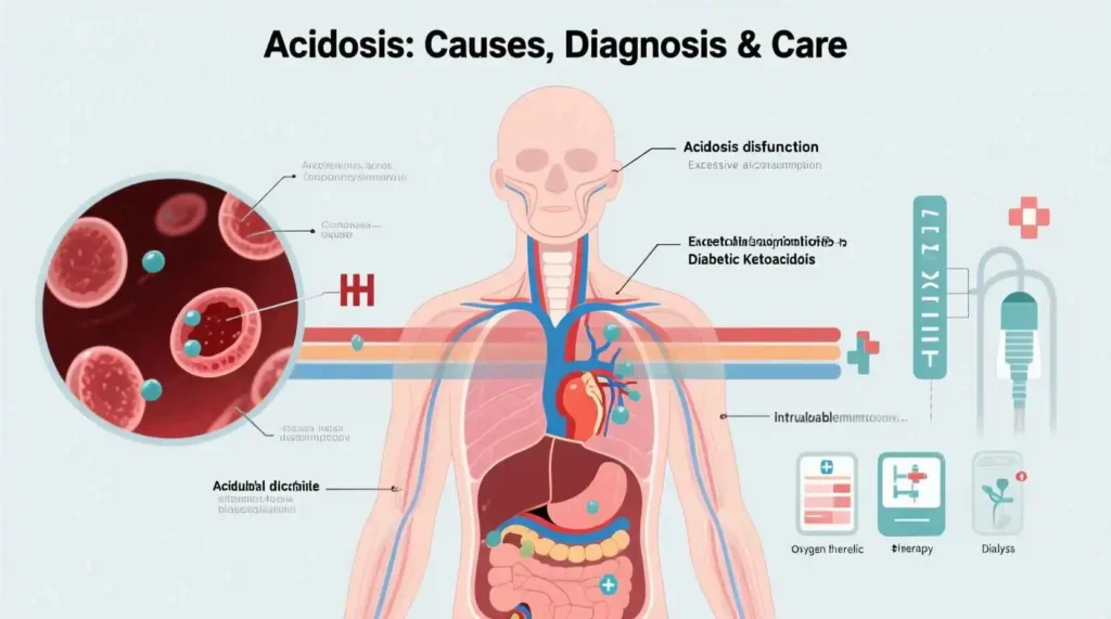
- Diabetic ketoacidosis (DKA): DKA occurs in patients with diabetes mellitus, typically type 1, when there is an absolute or relative insulin deficiency. Without sufficient insulin, glucose cannot enter cells for energy production, leading to increased lipolysis and the production of ketone bodies (acetoacetate, beta-hydroxybutyrate, and acetone). These ketone bodies are strong acids that accumulate in the blood, causing metabolic acidosis.
Example: A 25-year-old male with type 1 diabetes presents with nausea, vomiting, abdominal pain, and fruity-smelling breath. Laboratory tests reveal a blood glucose of 650 mg/dL, arterial pH of 7.15, bicarbonate of 10 mEq/L, and positive ketones in urine and blood. This is a classic presentation of DKA.
- Lactic acidosis: Lactic acidosis results from the accumulation of lactic acid, produced when cells undergo anaerobic metabolism due to inadequate oxygen delivery or utilization. Lactic acidosis is classified as either type A (due to tissue hypoperfusion and hypoxia) or type B (without evidence of tissue hypoxia).
Example A (Type A): A 65-year-old male with a history of coronary artery disease presents with severe chest pain and shortness of breath. He is found to be in cardiogenic shock with cool, clammy skin, blood pressure of 80/50 mmHg, and heart rate of 120 bpm. Laboratory tests reveal a lactate level of 8.2 mmol/L and arterial pH of 7.20. This represents type A lactic acidosis due to poor tissue perfusion from cardiogenic shock.
Example B (Type B): A 45-year-old female with a history of lymphoma presents with weakness and fatigue. She is currently receiving chemotherapy. Laboratory tests reveal a lactate level of 5.1 mmol/L and arterial pH of 7.28, despite normal blood pressure and oxygen saturation. This represents type B lactic acidosis, possibly related to her malignancy or chemotherapy.
- Uremia (chronic kidney disease): In advanced chronic kidney disease, the kidneys are unable to excrete hydrogen ions and regenerate bicarbonate, leading to metabolic acidosis. Additionally, the accumulation of organic acids such as phosphates, sulfates, and uric acid contributes to the high anion gap.
Example: A 70-year-old male with a history of hypertension and diabetes presents with fatigue, loss of appetite, and muscle cramps. Laboratory tests reveal a blood urea nitrogen (BUN) of 85 mg/dL, creatinine of 6.2 mg/dL, arterial pH of 7.22, bicarbonate of 12 mEq/L, and anion gap of 20 mEq/L. This represents metabolic acidosis due to uremia from end-stage renal disease.
- Toxin-induced acidosis: Several toxins can cause high anion gap metabolic acidosis through various mechanisms:
a. Methanol poisoning: Methanol is metabolized to formic acid, which inhibits mitochondrial respiration and causes metabolic acidosis. Symptoms include visual disturbances, CNS depression, and gastrointestinal complaints.
Example: A 40-year-old male presents with blurred vision, headache, and nausea after consuming what he believed to be ethanol. Laboratory tests reveal an anion gap of 25 mEq/L, arterial pH of 7.15, and an osmolar gap of 35 mOsm/kg. Serum methanol level is elevated, confirming methanol poisoning.
b. Ethylene glycol poisoning: Ethylene glycol is metabolized to glycolic acid and oxalic acid, which cause metabolic acidosis and can lead to crystalluria, acute kidney injury, and neurological symptoms.
Example: A 35-year-old male presents with altered mental status, nausea, and flank pain after ingesting antifreeze. Laboratory tests reveal an anion gap of 28 mEq/L, arterial pH of 7.10, and calcium oxalate crystals in the urine. Serum ethylene glycol level is elevated, confirming ethylene glycol poisoning.
c. Salicylate poisoning: Salicylates (aspirin) directly stimulate the respiratory center, causing respiratory alkalosis initially. With higher levels, salicylates uncouple oxidative phosphorylation and inhibit Krebs cycle enzymes, leading to lactic acid and ketoacid production.
Example: A 20-year-old female presents with tinnitus, hyperpnea, and confusion after an intentional aspirin overdose. Arterial blood gas reveals a pH of 7.45 (respiratory alkalosis) initially, but later tests show a pH of 7.20 (mixed respiratory alkalosis and metabolic acidosis). Serum salicylate level is 80 mg/dL.
- Other causes:
- Paraldehyde poisoning (rare in modern practice)
- Iron poisoning
- Isoniazid poisoning
- Toluene poisoning (can cause either high or normal anion gap acidosis)
Normal Anion Gap (Hyperchloremic) Metabolic Acidosis
Normal anion gap metabolic acidosis occurs when bicarbonate is lost from the body or when hydrogen ions are retained without an increase in unmeasured anions. In these cases, the decrease in bicarbonate is accompanied by an increase in chloride ions, maintaining a normal anion gap.
Common causes of normal anion gap metabolic acidosis include:
- Gastrointestinal bicarbonate loss:
- Diarrhea
- Pancreatic fistula
- Ureteral diversion
- Small bowel fistula
Example: A 30-year-old male presents with weakness, dizziness, and frequent watery stools for the past 3 days. He recently returned from a trip to a developing country. Laboratory tests reveal arterial pH of 7.25, bicarbonate of 15 mEq/L, and anion gap of 10 mEq/L. Stool studies confirm infection with Vibrio cholerae, causing bicarbonate loss through diarrhea.
- Renal tubular acidosis (RTA): Renal tubular acidosis refers to a group of disorders characterized by impaired renal acid excretion or bicarbonate reabsorption, leading to hyperchloremic metabolic acidosis. RTA is classified into several types:
a. Type 1 (distal) RTA: Characterized by impaired distal hydrogen ion secretion, resulting in inability to acidify urine below pH 5.5 despite systemic acidosis. It can be hereditary or acquired (e.g., autoimmune diseases, amphotericin B toxicity, sickle cell disease).
Example: A 25-year-old female with a history of Sjögren’s syndrome presents with fatigue, muscle weakness, and recurrent kidney stones. Laboratory tests reveal arterial pH of 7.28, bicarbonate of 16 mEq/L, potassium of 2.8 mEq/L, and urine pH of 6.8. These findings are consistent with type 1 RTA secondary to Sjögren’s syndrome.
b. Type 2 (proximal) RTA: Characterized by impaired proximal bicarbonate reabsorption, leading to bicarbonate wasting in the urine. It is often associated with other proximal tubular defects (Fanconi syndrome) and can be caused by multiple myeloma, heavy metal toxicity, or certain medications.
Example: A 40-year-old male with multiple myeloma presents with bone pain, fatigue, and muscle weakness. Laboratory tests reveal arterial pH of 7.30, bicarbonate of 17 mEq/L, and evidence of proximal tubular dysfunction including glycosuria and aminoaciduria. These findings are consistent with type 2 RTA due to multiple myeloma.
c. Type 4 RTA: Characterized by hypoaldosteronism or aldosterone resistance, leading to hyperkalemia and hyperchloremic metabolic acidosis. It is commonly associated with diabetes mellitus, chronic kidney disease, and medications that interfere with the renin-angiotensin-aldosterone system.
Example: A 60-year-old male with diabetes and hypertension presents with weakness and fatigue. He is taking lisinopril for his hypertension. Laboratory tests reveal arterial pH of 7.28, bicarbonate of 16 mEq/L, potassium of 5.8 mEq/L, and mildly elevated creatinine. These findings are consistent with type 4 RTA due to hyporeninemic hypoaldosteronism associated with diabetes and ACE inhibitor therapy.
- Early renal failure: In the early stages of chronic kidney disease, metabolic acidosis may be hyperchloremic due to impaired ammoniagenesis and reduced hydrogen ion secretion. As kidney disease progresses, the acidosis typically transitions to a high anion gap type.
- Carbonic anhydrase inhibitors: Medications such as acetazolamide inhibit carbonic anhydrase, reducing proximal bicarbonate reabsorption and leading to hyperchloremic metabolic acidosis.
Example: A 50-year-old female with glaucoma has been taking acetazolamide for several months. She presents with fatigue and tingling in her fingers and toes. Laboratory tests reveal arterial pH of 7.30, bicarbonate of 18 mEq/L, and anion gap of 10 mEq/L. These findings are consistent with acetazolamide-induced metabolic acidosis.
- Ureteral diversion: Surgical diversion of urine into the bowel (e.g., ileal conduit) can result in bicarbonate loss through the intestinal mucosa, leading to hyperchloremic metabolic acidosis.
- Acid administration: Infusion of large amounts of chloride-containing solutions (e.g., normal saline) or administration of acidifying agents (e.g., ammonium chloride, arginine hydrochloride) can cause hyperchloremic metabolic acidosis.
Example: A 55-year-old male undergoes major abdominal surgery and receives large volumes of normal saline during the procedure. Postoperatively, he develops metabolic acidosis with arterial pH of 7.28, bicarbonate of 17 mEq/L, and anion gap of 8 mEq/L. This represents dilutional acidosis due to excessive normal saline administration.
Mixed Acid-Base Disorders
