
A Comprehensive Guide to Diagnostic Tests: Types, Applications, and Interpretations
Introduction to Diagnostic Tests
Diagnostic tests form the cornerstone of modern medicine, serving as essential tools that healthcare professionals use to identify diseases, monitor health conditions, guide treatment decisions, and predict health outcomes. These tests range from simple physical examinations to complex molecular analyses, each providing valuable insights into the human body’s functioning and potential abnormalities. The evolution of diagnostic testing has revolutionized healthcare, enabling earlier detection of diseases, more accurate diagnoses, and personalized treatment approaches that have significantly improved patient outcomes across the globe.
The journey of diagnostic testing began centuries ago with basic observations of symptoms and physical signs, gradually progressing to more sophisticated techniques as scientific understanding advanced. Today, we have an unprecedented array of diagnostic tools at our disposal, each designed to answer specific clinical questions and provide objective data about a patient’s health status. From blood tests that reveal metabolic imbalances to imaging techniques that visualize internal structures, these tests collectively form a comprehensive framework for understanding human health and disease.
The importance of diagnostic tests cannot be overstated in contemporary healthcare. They serve multiple critical functions: screening for diseases in asymptomatic individuals, confirming or ruling out suspected conditions, assessing disease severity, monitoring treatment effectiveness, and predicting prognosis. Without accurate diagnostic tests, medical decision-making would rely heavily on subjective assessments and guesswork, potentially leading to delayed treatments, inappropriate therapies, and poorer patient outcomes.

As medical science continues to advance, the field of diagnostic testing evolves at a remarkable pace. New technologies emerge regularly, offering improved accuracy, faster results, less invasive procedures, and more comprehensive information. These advancements are transforming how we approach disease detection and management, shifting the paradigm from reactive treatment to proactive prevention and personalized medicine. Understanding the various types of diagnostic tests, their applications, and their limitations is essential for both healthcare professionals and patients navigating the complex landscape of modern healthcare.
This comprehensive guide explores the diverse world of diagnostic testing, examining the major categories of tests, their principles and applications, and their role in contemporary healthcare. We will delve into the specifics of common diagnostic procedures, discuss recent advancements in the field, and address frequently asked questions to provide a thorough understanding of this critical aspect of medical practice.
Categories of Diagnostic Tests
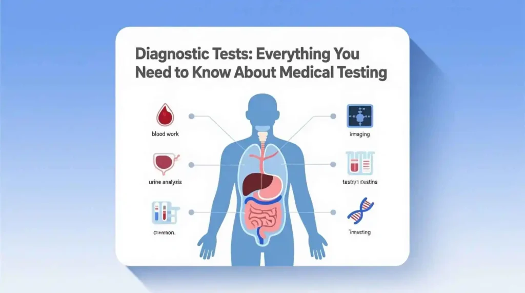
Diagnostic tests can be broadly categorized based on the type of information they provide, the techniques they employ, or the biological samples they analyze. Understanding these categories helps healthcare providers select the most appropriate tests for specific clinical scenarios and enables patients to better comprehend their diagnostic journeys. The major categories of diagnostic tests include laboratory tests, imaging tests, genetic tests, physiological tests, and pathological tests, each encompassing a wide range of specific procedures designed to answer different clinical questions.
Laboratory Tests
Laboratory tests analyze biological specimens such as blood, urine, cerebrospinal fluid, or tissue samples to detect and measure various substances, cells, or microorganisms. These tests provide quantitative and qualitative information about biochemical processes, organ function, infections, and other physiological parameters. Laboratory medicine is one of the most frequently used diagnostic categories, with billions of tests performed annually worldwide.
Blood tests are perhaps the most common type of laboratory investigation, encompassing numerous specific analyses. A complete blood count (CBC) measures the quantity and characteristics of different blood cells, providing information about conditions like anemia, infection, and certain cancers. Blood chemistry panels assess electrolytes, kidney function, liver function, blood glucose, and lipid levels, offering insights into metabolic health and organ function. Specialized blood tests can detect hormones, antibodies, tumor markers, and specific proteins associated with various diseases.
Urine tests analyze urine samples for abnormalities in composition, presence of cells, microorganisms, or substances that shouldn’t normally be present. Urinalysis can detect urinary tract infections, kidney diseases, metabolic disorders, and systemic conditions like diabetes. Similarly, cerebrospinal fluid analysis examines the fluid surrounding the brain and spinal cord to diagnose infections, inflammatory conditions, and certain neurological disorders.
Microbiological tests identify and characterize microorganisms such as bacteria, viruses, fungi, and parasites in clinical specimens. These tests include cultures, microscopic examinations, antigen detection, and molecular techniques like polymerase chain reaction (PCR). Microbiological testing is crucial for diagnosing infectious diseases and guiding antimicrobial therapy.
Imaging Tests
Imaging tests create visual representations of internal body structures to detect abnormalities, assess organ function, and guide medical procedures. These non-invasive or minimally invasive techniques have revolutionized medical diagnostics by allowing healthcare providers to visualize anatomical structures and physiological processes without surgery.
X-ray imaging uses electromagnetic radiation to create images of bones, chest structures, and certain soft tissues. It’s commonly used to detect fractures, pneumonia, and certain tumors. Computed tomography (CT) scans combine X-ray technology with computer processing to create detailed cross-sectional images of the body, providing more detailed information than conventional X-rays. CT scans are particularly valuable for evaluating trauma, detecting tumors, and assessing vascular conditions.
Magnetic resonance imaging (MRI) uses powerful magnets and radio waves to generate detailed images of soft tissues, including the brain, spinal cord, muscles, and joints. MRI is especially useful for neurological conditions, musculoskeletal disorders, and certain cancers. Ultrasound imaging employs high-frequency sound waves to create real-time images of internal structures, making it valuable for examining the heart, blood vessels, abdominal organs, and during pregnancy.
Nuclear medicine imaging involves administering small amounts of radioactive substances that accumulate in specific organs or tissues, allowing visualization of metabolic processes using special cameras. Positron emission tomography (PET) scans, often combined with CT (PET-CT), are particularly useful for detecting cancer, evaluating heart disease, and assessing brain function.
Genetic Tests
Genetic tests analyze DNA, RNA, chromosomes, or proteins to identify genetic variations associated with inherited disorders, disease susceptibility, or pharmacogenomic responses. These tests have become increasingly important with the growing understanding of the role of genetics in health and disease.
Chromosomal analysis examines the number and structure of chromosomes to detect abnormalities like Down syndrome, Turner syndrome, and certain leukemias. DNA sequencing identifies specific changes in DNA sequences that cause or predispose to genetic disorders such as cystic fibrosis, sickle cell disease, and hereditary cancer syndromes. Biochemical genetic tests measure the activity or amount of specific proteins that indicate genetic disorders, such as phenylketonuria (PKU) or Tay-Sachs disease.
Pharmacogenomic tests analyze how genetic variations affect an individual’s response to medications, helping to guide drug selection and dosing. These tests are increasingly used in oncology, psychiatry, and cardiology to personalize treatment approaches and minimize adverse drug reactions.
Physiological Tests
Physiological tests evaluate the function of specific organs or body systems by measuring their performance under controlled conditions. These tests provide dynamic information about how the body responds to stress, activity, or other stimuli, complementing the static information provided by laboratory and imaging tests.
Electrocardiography (ECG or EKG) records the electrical activity of the heart to detect arrhythmias, heart attacks, and other cardiac conditions. Stress testing evaluates heart function during physical exertion, often using treadmill exercise or pharmacological agents to simulate exercise. Pulmonary function tests measure lung capacity, airflow, and gas exchange to diagnose and monitor respiratory conditions like asthma and chronic obstructive pulmonary disease (COPD).
Electroencephalography (EEG) records electrical activity in the brain to help diagnose epilepsy, sleep disorders, and certain neurological conditions. Electromyography (EMG) assesses muscle function and nerve conduction to diagnose neuromuscular disorders. Nerve conduction studies measure how well electrical signals travel through nerves, helping to identify nerve damage or dysfunction.
Pathological Tests
Pathological tests involve the examination of tissues and cells to diagnose diseases, particularly cancer. These tests provide definitive diagnoses by allowing direct visualization of cellular abnormalities under a microscope.
Biopsy procedures involve removing small samples of tissue from suspicious areas for microscopic examination. Different types of biopsies include needle biopsies, endoscopic biopsies, excisional biopsies, and surgical biopsies, each appropriate for different clinical scenarios. Cytology examines individual cells or small clusters of cells obtained from body fluids, fine-needle aspirations, or exfoliative samples like Pap smears.
Histopathology involves processing tissue samples, embedding them in paraffin, cutting thin sections, staining them, and examining them under a microscope. This technique is the gold standard for diagnosing cancer and many other diseases. Immunohistochemistry uses antibodies to detect specific proteins in tissue sections, helping to classify tumors and guide treatment decisions. Molecular pathology analyzes DNA, RNA, or proteins in tissue samples to identify genetic alterations that inform diagnosis, prognosis, and treatment selection.
Common Laboratory Tests and Their Applications
Laboratory testing constitutes a significant portion of diagnostic procedures performed in healthcare settings, providing essential information about a patient’s biochemical and physiological status. These tests analyze various biological specimens to detect abnormalities, monitor disease progression, and assess treatment responses. Understanding the most common laboratory tests, their principles, and clinical applications is fundamental for healthcare providers and patients alike.
Complete Blood Count (CBC)
The complete blood count is one of the most frequently performed laboratory tests, providing comprehensive information about the cellular components of blood. A CBC measures several parameters:
- Red blood cells (RBCs): These cells carry oxygen from the lungs to tissues and carbon dioxide back to the lungs. The test measures the number of RBCs, their size (mean corpuscular volume, MCV), the amount of hemoglobin (the oxygen-carrying protein), and the proportion of blood made up by RBCs (hematocrit).
- White blood cells (WBCs): These cells protect the body against infection. An elevated count may indicate infection, inflammation, or leukemia, while a decreased count may signal bone marrow problems or certain infections.
- Platelets: These small cell fragments help blood to clot. Abnormal platelet counts can indicate bleeding disorders or bone marrow diseases.
The CBC is used to help diagnose conditions such as anemia, infection, and certain cancers. It’s also used to monitor patients undergoing treatments that affect blood cell production, such as chemotherapy.
Comprehensive Metabolic Panel (CMP)
The comprehensive metabolic panel is a group of blood tests that provides information about your body’s chemical balance and metabolism. It typically includes:
- Glucose: Elevated levels may indicate diabetes, while low levels can signal hypoglycemia.
- Calcium: Abnormal levels can indicate problems with bones, kidneys, or parathyroid glands.
- Electrolytes: Including sodium, potassium, chloride, and bicarbonate, which help regulate fluid balance, nerve function, and muscle contraction.
- Kidney function tests: Blood urea nitrogen (BUN) and creatinine levels indicate how well the kidneys are filtering waste from the blood.
- Liver function tests: Including albumin, total protein, alkaline phosphatase (ALP), alanine aminotransferase (ALT), aspartate aminotransferase (AST), and bilirubin, which assess liver health and function.
The CMP is commonly used to screen for conditions like diabetes, kidney disease, and liver disease. It’s also used to monitor patients with known conditions and to check the effects of medications that might affect kidney or liver function.
Lipid Panel
The lipid panel measures fats and fatty substances in the blood, which are important indicators of cardiovascular health. The test typically includes:
- Total cholesterol
- Low-density lipoprotein (LDL) cholesterol, often called “bad” cholesterol
- High-density lipoprotein (HDL) cholesterol, often called “good” cholesterol
- Triglycerides, a type of fat in the blood
Abnormal lipid levels are associated with an increased risk of atherosclerosis, heart attack, and stroke. The lipid panel is used to assess cardiovascular risk, guide treatment decisions, and monitor the effectiveness of cholesterol-lowering medications.
Thyroid Function Tests
Thyroid function tests evaluate how well the thyroid gland is working by measuring levels of thyroid hormones and thyroid-stimulating hormone (TSH). Common tests include:
- Thyroid-stimulating hormone (TSH): Produced by the pituitary gland, TSH regulates thyroid hormone production. Abnormal levels can indicate hypothyroidism (underactive thyroid) or hyperthyroidism (overactive thyroid).
- Thyroxine (T4): The main hormone produced by the thyroid gland. Most T4 is bound to proteins, but free T4 is the biologically active form.
- Triiodothyronine (T3): Another thyroid hormone, mostly produced by conversion from T4 in peripheral tissues. Free T3 is the active form.
Thyroid function tests are used to diagnose thyroid disorders, monitor treatment for thyroid conditions, and investigate symptoms like fatigue, weight changes, and mood disturbances.
Cardiac Biomarkers
Cardiac biomarkers are substances released into the blood when the heart is damaged or stressed. These tests are crucial for diagnosing and managing heart conditions. Common cardiac biomarkers include:
- Troponins: Proteins found in heart muscle that are released into the blood when the heart is damaged. Elevated troponin levels are highly specific for heart muscle injury and are used to diagnose heart attacks.
- Creatine kinase-MB (CK-MB): An enzyme found in heart muscle that increases with heart damage. It’s less specific than troponin but can help determine the timing of a heart attack.
- B-type natriuretic peptide (BNP) or N-terminal pro-BNP (NT-proBNP): Hormones released when the heart is stretched, as in heart failure. Elevated levels help diagnose and assess the severity of heart failure.
Cardiac biomarkers are essential in emergency settings for evaluating chest pain, diagnosing heart attacks, and assessing heart failure. They’re also used to monitor treatment effectiveness and predict prognosis in cardiac patients.
Liver Function Tests
Liver function tests (LFTs) measure various enzymes, proteins, and substances produced or processed by the liver. These tests help assess liver health and detect liver damage or disease. Common LFTs include:
- Alanine aminotransferase (ALT): An enzyme found mainly in the liver. Elevated levels indicate liver cell damage.
- Aspartate aminotransferase (AST): An enzyme found in the liver and other tissues. Elevated levels can indicate liver damage but are less specific than ALT.
- Alkaline phosphatase (ALP): An enzyme found in the liver and bones. Elevated levels can indicate liver disease or bone disorders.
- Bilirubin: A waste product processed by the liver. Elevated levels can indicate liver disease or certain types of anemia.
- Albumin and total protein: Proteins produced by the liver. Low levels can indicate chronic liver disease.
Liver function tests are used to diagnose liver diseases like hepatitis and cirrhosis, monitor the progression of liver disease, assess the effects of medications that might affect the liver, and evaluate liver function before surgery.
Kidney Function Tests
Kidney function tests evaluate how well the kidneys are filtering waste from the blood and maintaining fluid and electrolyte balance. Common tests include:
- Blood urea nitrogen (BUN): A waste product filtered by the kidneys. Elevated levels can indicate kidney dysfunction or dehydration.
- Creatinine: A waste product from muscle metabolism that’s filtered by the kidneys. Elevated levels indicate decreased kidney function.
- Glomerular filtration rate (GFR): A calculation based on creatinine levels, age, sex, and race that estimates how well the kidneys are filtering waste. It’s the best overall indicator of kidney function.
- Urinalysis: Examination of urine for protein, blood, and other substances that can indicate kidney disease.
Kidney function tests are used to diagnose and monitor kidney disease, assess the effects of medications on kidney function, and evaluate kidney function before certain medical procedures or surgeries.
Common Laboratory Tests and Their Clinical Applications
| Test Category | Specific Test | Measures | Clinical Applications |
| Hematology | Complete Blood Count (CBC) | Red blood cells, white blood cells, platelets | Anemia, infection, leukemia, monitoring chemotherapy |
| Chemistry | Comprehensive Metabolic Panel (CMP) | Glucose, electrolytes, kidney and liver function markers | Diabetes, kidney disease, liver disease, metabolic disorders |
| Chemistry | Lipid Panel | Total cholesterol, LDL, HDL, triglycerides | Cardiovascular risk assessment, monitoring lipid-lowering therapy |
| Endocrinology | Thyroid Function Tests | TSH, T4, T3 | Hypothyroidism, hyperthyroidism, thyroid disorders |
| Cardiology | Cardiac Biomarkers | Troponins, CK-MB, BNP/NT-proBNP | Heart attack diagnosis, heart failure assessment, cardiac risk stratification |
| Hepatology | Liver Function Tests | ALT, AST, ALP, bilirubin, albumin | Hepatitis, cirrhosis, liver damage, medication monitoring |
| Nephrology | Kidney Function Tests | BUN, creatinine, GFR, urinalysis | Kidney disease, renal function monitoring, fluid balance assessment |
| Immunology | C-Reactive Protein (CRP) | Inflammation marker | Infection, inflammatory conditions, cardiovascular risk |
| Hematology | Coagulation Panel | PT, INR, PTT, fibrinogen | Bleeding disorders, anticoagulant monitoring, thrombosis risk |
| Microbiology | Blood Culture | Bacteria or fungi in blood | Sepsis, bloodstream infections, fever of unknown origin |
Imaging Tests: Principles and Applications
Imaging tests have transformed medical diagnostics by allowing healthcare providers to visualize internal structures and processes without invasive procedures. These techniques use various forms of energy to create detailed images of the body, providing invaluable information about anatomy, physiology, and pathology. From simple X-rays to complex molecular imaging, each modality offers unique advantages for specific clinical applications.
X-Ray Imaging
X-ray imaging is one of the oldest and most widely used diagnostic techniques, utilizing electromagnetic radiation to penetrate the body and create images of internal structures. The principle behind X-ray imaging is that different tissues absorb X-rays to varying degrees based on their density and composition. Dense tissues like bones absorb more X-rays and appear white on the image, while soft tissues appear in shades of gray, and air appears black.
Conventional X-ray imaging is commonly used to examine bones for fractures, dislocations, and infections. It’s also frequently employed to evaluate the chest for pneumonia, lung tumors, heart size, and pleural effusions. Abdominal X-rays can detect intestinal obstructions, kidney stones, and certain types of tumors. Dental X-rays are essential for identifying cavities, impacted teeth, and jawbone problems.
Contrast agents are sometimes used with X-ray imaging to enhance visualization of specific structures. For example, barium sulfate is used to outline the gastrointestinal tract in upper GI series and barium enemas, while iodinated contrast agents are used to visualize blood vessels in angiography and the urinary tract in intravenous pyelograms.
Despite its widespread use, X-ray imaging has limitations. It provides two-dimensional images that can sometimes obscure details, and it has relatively poor soft tissue contrast compared to other imaging modalities. Additionally, X-rays involve ionizing radiation, which carries potential risks, particularly with repeated exposure.
Computed Tomography (CT)
Computed tomography, commonly known as CT or CAT scan, is an advanced imaging technique that combines X-ray technology with computer processing to create detailed cross-sectional images of the body. During a CT scan, an X-ray tube rotates around the patient, capturing multiple images from different angles. A computer then processes these images to generate detailed cross-sectional views, which can be further reconstructed into three-dimensional representations.
CT imaging offers several advantages over conventional X-rays. It provides much greater detail and contrast, allowing for better visualization of soft tissues, blood vessels, and small abnormalities. CT scans are particularly valuable for evaluating trauma patients, as they can quickly detect internal injuries, bleeding, and fractures. They’re also commonly used to diagnose and stage cancers, assess vascular diseases like aneurysms and pulmonary embolism, and guide biopsies and other procedures.
Modern CT scanners have evolved significantly, with multi-detector CT (MDCT) allowing for faster scanning with higher resolution. Dual-energy CT uses two different X-ray energies to provide additional information about tissue composition, while CT angiography combines CT scanning with contrast agents to create detailed images of blood vessels.
Despite its diagnostic capabilities, CT scanning involves higher radiation exposure than conventional X-rays, raising concerns about cumulative radiation dose, particularly in children and young adults. Additionally, the use of iodinated contrast agents can cause allergic reactions in some patients and may pose risks for individuals with kidney problems.
Magnetic Resonance Imaging (MRI)
Magnetic resonance imaging is a powerful diagnostic technique that uses strong magnetic fields and radio waves to generate detailed images of soft tissues in the body. Unlike X-rays and CT scans, MRI does not use ionizing radiation, making it safer for repeated examinations, particularly in children and pregnant women.
The principle behind MRI is based on the behavior of hydrogen atoms (primarily in water molecules) in a magnetic field. When exposed to radio waves, these atoms emit signals that are detected by the MRI scanner and processed by computers to create detailed images. Different tissues emit different signals based on their water content and molecular environment, allowing for excellent soft tissue contrast.
MRI is particularly valuable for imaging the brain, spinal cord, joints, muscles, and other soft tissues. It’s commonly used to diagnose neurological conditions like multiple sclerosis, brain tumors, and spinal cord injuries. In orthopedics, MRI is the gold standard for evaluating ligament and tendon injuries, cartilage damage, and bone marrow abnormalities. Cardiac MRI provides detailed information about heart structure and function, while breast MRI is used as an adjunct to mammography in high-risk patients.
Specialized MRI techniques provide additional diagnostic information. Functional MRI (fMRI) measures brain activity by detecting changes in blood flow, making it valuable for planning brain surgery and studying neurological disorders. Diffusion tensor imaging (DTI) maps the brain’s white matter tracts, helping to assess conditions like traumatic brain injury and stroke. Magnetic resonance angiography (MRA) visualizes blood vessels without the need for contrast agents in some cases.
While MRI offers excellent image quality and safety advantages, it has limitations. The scanners are expensive and not as widely available as CT or X-ray machines. MRI examinations take longer than other imaging tests, and the strong magnetic field requires careful screening for metal implants or devices in patients. Some patients experience claustrophobia during MRI scans, and the loud noise generated by the scanner can be uncomfortable.
Ultrasound Imaging
Ultrasound imaging, also known as sonography, uses high-frequency sound waves to create real-time images of internal structures. During an ultrasound examination, a transducer emits sound waves into the body, and the echoes reflected back from tissues are detected and converted into images by a computer.
Ultrasound imaging has several advantages that make it widely used in clinical practice. It’s non-invasive, does not use ionizing radiation, is relatively inexpensive, and provides real-time imaging, allowing for dynamic assessment of moving structures like the heart and blood vessels. Ultrasound is also portable, making it valuable for bedside examinations and in resource-limited settings.
Obstetric ultrasound is one of the most common applications, used to monitor fetal development during pregnancy. In cardiology, echocardiography evaluates heart structure and function, including valve function, chamber sizes, and blood flow. Abdominal ultrasound examines organs like the liver, gallbladder, kidneys, pancreas, and spleen, helping to detect gallstones, kidney stones, tumors, and cysts. Doppler ultrasound assesses blood flow in vessels, aiding in the diagnosis of conditions like deep vein thrombosis and peripheral artery disease.
Recent advancements in ultrasound technology have expanded its capabilities. Contrast-enhanced ultrasound uses microbubble contrast agents to improve visualization of blood flow and tissue perfusion. Elastography measures tissue stiffness, helping to differentiate between benign and malignant lesions in organs like the liver and breast. Three-dimensional and four-dimensional (3D/4D) ultrasound provides more detailed images, particularly useful in obstetrics and cardiology.
Despite its many advantages, ultrasound has limitations. Image quality can be affected by patient body habitus, with obesity often reducing image clarity. Ultrasound waves cannot penetrate bone or gas, limiting visualization of structures behind bones or in the bowel. Additionally, ultrasound imaging is highly operator-dependent, requiring skilled technicians for optimal results.
Nuclear Medicine Imaging
Nuclear medicine imaging involves administering small amounts of radioactive substances (radiopharmaceuticals) that accumulate in specific organs or tissues. Special cameras detect the gamma rays emitted by these substances, creating images that show how organs and tissues are functioning.
Unlike other imaging modalities that primarily show anatomy, nuclear medicine provides functional information about physiological processes at the molecular level. This makes it particularly valuable for detecting diseases at their earliest stages, often before structural changes become apparent.
Common nuclear medicine procedures include bone scans, which detect cancer that has spread to the bones; thyroid scans, which evaluate thyroid function and detect nodules; and cardiac perfusion scans, which assess blood flow to the heart muscle. Positron emission tomography (PET) scans are increasingly used in oncology to detect cancer, assess treatment response, and determine if cancer has spread. PET scans are often combined with CT scans (PET-CT) to provide both functional and anatomical information in a single examination.
Single-photon emission computed tomography (SPECT) is another nuclear medicine technique that provides three-dimensional images of organ function. SPECT is commonly used in cardiology for myocardial perfusion imaging, in neurology for brain function studies, and in bone scanning.
While nuclear medicine imaging provides unique functional information, it involves exposure to radiation, though typically at levels comparable to or lower than CT scans. The radioactive substances used have short half-lives and are eliminated from the body relatively quickly. Nuclear medicine also requires specialized facilities and expertise, limiting its availability in some settings.
Comparison of Imaging Modalities
| Imaging Modality | Physical Principle | Primary Clinical Applications | Advantages | Limitations |
| X-Ray | Electromagnetic radiation | Bone fractures, chest imaging, dental exams | Fast, widely available, inexpensive | Limited soft tissue contrast, 2D images, ionizing radiation |
| CT | X-rays with computer processing | Trauma, cancer staging, vascular imaging | Detailed cross-sectional images, fast, good for acute conditions | Higher radiation dose, contrast agent risks, less soft tissue contrast than MRI |
| MRI | Magnetic fields and radio waves | Brain, spinal cord, joints, soft tissues | Excellent soft tissue contrast, no ionizing radiation, multiplanar imaging | Expensive, longer scan times, contraindicated with certain implants, claustrophobia |
| Ultrasound | High-frequency sound waves | Obstetrics, cardiology, abdominal organs, blood flow | Real-time imaging, no radiation, portable, inexpensive | Operator-dependent, limited by bone and gas, reduced quality in obesity |
| Nuclear Medicine | Radioactive tracers | Cancer detection, thyroid function, cardiac perfusion | Functional information, detects disease at molecular level | Radiation exposure, limited availability, lower spatial resolution |
Genetic and Molecular Diagnostics
The field of genetic and molecular diagnostics has expanded exponentially in recent years, revolutionizing our understanding of disease mechanisms and enabling personalized approaches to healthcare. These tests analyze DNA, RNA, chromosomes, proteins, and metabolites to detect genetic variations, mutations, and molecular alterations associated with diseases. By examining the fundamental building blocks of life, genetic and molecular diagnostics provide insights that were unimaginable just a few decades ago, transforming how we diagnose, treat, and prevent diseases.
Chromosomal Analysis
Chromosomal analysis, also known as karyotyping, examines the number and structure of chromosomes within cells. Humans normally have 46 chromosomes arranged in 23 pairs, with one set inherited from each parent. Chromosomal abnormalities can lead to a variety of genetic disorders and developmental problems.
Traditional karyotyping involves staining chromosomes and viewing them under a microscope to identify numerical abnormalities (such as trisomy 21 in Down syndrome) and large structural abnormalities (such as translocations, deletions, or duplications). This technique is commonly used in prenatal diagnosis, particularly for older pregnant women or those with abnormal screening results. It’s also used to diagnose genetic syndromes in children with developmental delays or congenital abnormalities, and in cases of infertility or recurrent miscarriages.
Fluorescence in situ hybridization (FISH) is a more targeted approach that uses fluorescent DNA probes to bind to specific chromosome regions. FISH can detect smaller chromosomal abnormalities that might be missed by traditional karyotyping, such as microdeletions or microduplications. It’s particularly valuable in diagnosing conditions like DiGeorge syndrome (22q11.2 deletion syndrome) and certain cancers where specific chromosomal translocations are characteristic.
Chromosomal microarray analysis (CMA) represents a more advanced technique that can detect smaller chromosomal imbalances than traditional karyotyping or FISH. CMA uses microchips containing thousands of DNA probes to screen the entire genome for copy number variations (CNVs), including deletions and duplications too small to be seen with a microscope. This technique has become a first-tier test for children with developmental delays, autism spectrum disorders, or multiple congenital anomalies.
DNA Sequencing
DNA sequencing determines the precise order of nucleotides (adenine, thymine, cytosine, and guanine) in a DNA molecule, allowing for the identification of genetic variations that may cause or predispose to disease. Several sequencing technologies are used in clinical diagnostics, each with different applications and capabilities.
Sanger sequencing, developed in the 1970s, was the first method to determine the sequence of DNA fragments. While largely replaced by newer technologies for large-scale sequencing, Sanger sequencing remains valuable for confirming specific genetic variants identified by other methods and for analyzing individual genes or small gene panels.
Next-generation sequencing (NGS) technologies have revolutionized genetic testing by allowing for the simultaneous sequencing of millions of DNA fragments. NGS can be applied in various ways:
- Targeted gene panels: Sequence specific genes known to be associated with particular conditions, such as hereditary cancer syndromes or cardiomyopathies. This approach is cost-effective and efficient when there’s a strong suspicion of a specific genetic disorder.
- Whole exome sequencing (WES): Sequences the protein-coding regions of the genome (the exome), which harbors approximately 85% of known disease-causing mutations. WES is particularly useful for patients with complex or undiagnosed genetic disorders.
- Whole genome sequencing (WGS): Sequences the entire genome, including both coding and non-coding regions. While more expensive and generating more data than WES, WGS provides the most comprehensive genetic information and is increasingly used in research and clinical diagnostics.
DNA sequencing has numerous clinical applications. In oncology, it identifies somatic mutations in tumors that can guide targeted therapy decisions. In medical genetics, it diagnoses rare genetic disorders and enables carrier screening for recessive conditions. In pharmacogenomics, it identifies genetic variations that affect drug metabolism and response, helping to personalize medication choices.
Biochemical Genetic Testing
Biochemical genetic testing measures the activity or amount of specific proteins or metabolites to detect inborn errors of metabolism. These tests are particularly valuable for diagnosing disorders that may not be apparent through DNA sequencing alone, such as those affecting enzyme function or metabolic pathways.
Newborn screening programs use biochemical tests to detect certain genetic disorders shortly after birth, allowing for early intervention that can prevent severe complications. Common conditions screened for include phenylketonuria (PKU), congenital hypothyroidism, sickle cell disease, and maple syrup urine disease. These tests typically analyze dried blood spots collected from a heel prick shortly after birth.
Quantitative amino acid analysis measures levels of amino acids in blood or urine to detect disorders of amino acid metabolism, such as PKU or homocystinuria. Organic acid analysis identifies abnormal organic acids in urine that accumulate in disorders of organic acid metabolism, such as methylmalonic acidemia. Enzyme activity assays measure the function of specific enzymes, helping to diagnose lysosomal storage diseases like Gaucher disease or Tay-Sachs disease.
Tandem mass spectrometry (MS/MS) has become a cornerstone of biochemical genetic testing, allowing for the simultaneous analysis of multiple metabolites from a single sample. This technology has expanded newborn screening programs and improved the diagnosis of metabolic disorders.
Molecular Infectious Disease Testing
Molecular techniques have transformed the diagnosis of infectious diseases by providing rapid, sensitive, and specific detection of pathogens. These methods detect nucleic acids (DNA or RNA) from viruses, bacteria, fungi, or parasites, often before traditional methods like culture or serology can identify the infection.
Polymerase chain reaction (PCR) is the most widely used molecular technique for infectious disease diagnosis. PCR amplifies specific DNA sequences, allowing for the detection of even small amounts of pathogen genetic material. Real-time PCR (qPCR) combines amplification with detection, providing quantitative results and faster turnaround times. PCR is used to diagnose a wide range of infections, including HIV, hepatitis C, influenza, tuberculosis, and sexually transmitted infections.
Nucleic acid amplification tests (NAATs) encompass various techniques similar to PCR that amplify and detect pathogen nucleic acids. These tests have become the gold standard for many infections due to their high sensitivity and specificity. For example, NAATs are the preferred method for diagnosing chlamydia and gonorrhea, and they’re increasingly used for rapid detection of respiratory viruses.
Multiplex PCR panels can detect multiple pathogens simultaneously from a single sample, making them particularly valuable for syndromic testing where multiple pathogens can cause similar symptoms. For example, respiratory panels can detect dozens of viruses and bacteria that cause respiratory infections, while gastrointestinal panels can identify various pathogens causing diarrhea.
Next-generation sequencing is increasingly used in infectious disease diagnostics, particularly for identifying unknown or unexpected pathogens. Metagenomic NGS sequences all nucleic acids in a sample, allowing for the detection of any pathogen present without prior suspicion of what it might be. This approach is valuable for diagnosing encephalitis, sepsis, and other complex infectious conditions.
Pharmacogenomic Testing