

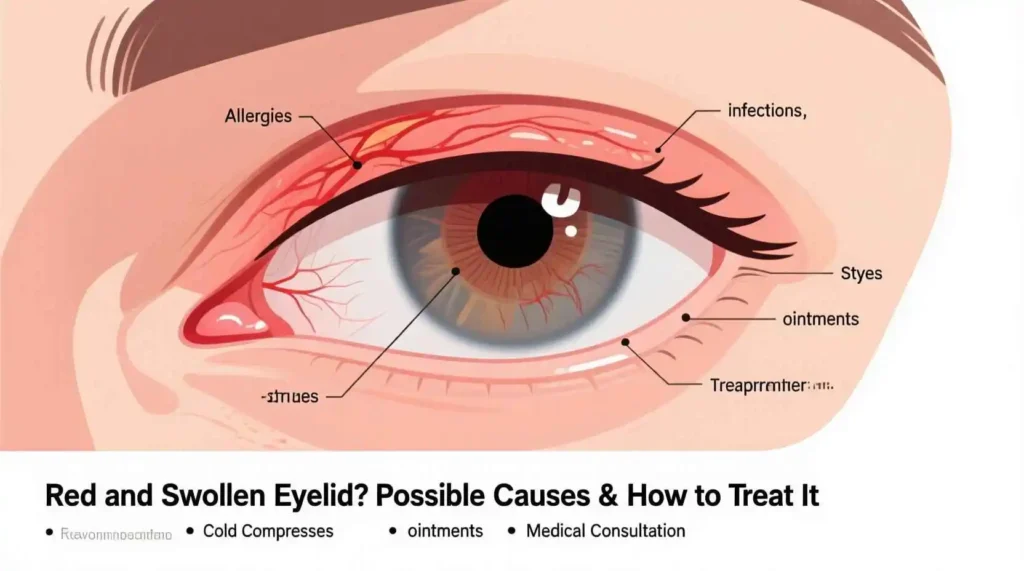
Red and Swollen Eyelid? Possible Causes & How to Treat It
“The eyes are the windows to the soul, but they are also the most exposed and vulnerable organs of the body. Prompt recognition of subtle changes can prevent serious complications.” — Dr. Jane L. Patel, MD, Ophthalmology, American Academy of Ophthalmology
Introduction
When we notice a red, swollen upper eyelid that has crept up slowly over days or weeks, accompanied by mild tearing and a stinging sensation each time we blink, it is natural to feel uneasy. The eyelid is a delicate structure, and even minor disturbances can affect vision, comfort, and overall eye health. In this article we will explore:
- The most common causes of this presentation.
- How clinicians diagnose the underlying problem.
- Evidence‑based treatment options and self‑care measures.
Our goal is to equip you with a clear, step‑by‑step understanding so you can recognize red flags, seek appropriate care, and manage the condition effectively.

Anatomy Refresher: Why the Upper Eyelid Matters
| Structure | Function | Relevance to Swelling |
| Skin & Sebaceous Glands | Protects the eye; produces oily tear film | Blockage → stye or chalazion |
| Orbicularis Oculi Muscle | Closes eyelid; distributes tears | Inflammation → pain on blinking |
| Meibomian Glands | Secrete lipid layer of tears | Dysfunction leads to dry eye & swelling |
| Conjunctiva | Thin mucous membrane lining eyelid | Conjunctivitis manifests as redness |
| Lacrimal Gland & Ducts | Produce aqueous tear component | Obstruction → watery discharge |
Understanding these layers helps us pinpoint where a problem might arise when the eyelid looks inflamed.
Common Causes of Gradual Upper‑Lid Swelling
Below is a concise list of the most frequent etiologies, ranked by likelihood in otherwise healthy adults.
- Stye (Hordeolum) – Acute, bacterial infection of a glandular opening
- Chalazion – Chronic blockage of a Meibomian gland
- Blepharitis – Inflammation of the eyelid margin
- Allergic Conjunctivitis – Seasonal or environmental allergen exposure
- Contact Dermatitis – Irritation from cosmetics, eye drops, or topical meds
- Preseptal (Periorbital) Cellulitis – Bacterial infection of the eyelid and skin
- Dacryocystitis – Lacrimal sac infection that can cause secondary lid swelling
- Orbital Tumors (rare) – Benign or malignant masses causing progressive lid thickening
Gradual upper lid swelling is a common occurrence in otherwise healthy adults, and it can be attributed to several underlying causes. The most frequent etiologies, ranked by likelihood, are as follows:
Stye (Hordeolum): A stye is an acute bacterial infection that affects a glandular opening on the eyelid, usually resulting from the blockage of an oil gland called the meibomian gland. The infection causes a painful, red, and swollen bump on the eyelid, which can lead to gradual upper lid swelling.
Chalazion: A chalazion is a chronic blockage of a meibomian gland, which can result in a small, painless lump on the upper or lower eyelid. This blockage occurs when the oil produced by the gland thickens and hardens, causing the gland to become clogged. The swelling can gradually worsen if left untreated.
Blepharitis: This condition involves inflammation of the eyelid margin, often caused by an overgrowth of bacteria or a malfunction of the oil glands in the eyelids. Blepharitis can lead to gradual upper lid swelling, as well as symptoms such as itching, redness, and crusting of the eyelids.
Allergic Conjunctivitis: This is an inflammation of the conjunctiva, the clear tissue that covers the white part of the eye and the inside of the eyelids. It is usually triggered by exposure to seasonal or environmental allergens, such as pollen or pet dander. Allergic conjunctivitis can cause gradual upper lid swelling, as well as symptoms such as itching, redness, and watery eyes.
Contact Dermatitis: This is an irritation of the skin caused by contact with cosmetics, eye drops, or topical medications. The irritation can lead to gradual upper lid swelling, as well as symptoms such as redness, itching, and a burning sensation.
Preseptal (Periorbital) Cellulitis: This is a bacterial infection of the eyelid and skin surrounding the eye. It can cause gradual upper lid swelling, along with symptoms such as redness, warmth, and tenderness of the affected area. Preseptal cellulitis typically requires antibiotic treatment to prevent the infection from spreading to the deeper tissues of the eye.
Dacryocystitis: This is an infection of the lacrimal sac, a small gland located near the inner corner of the eye. The infection can cause gradual upper lid swelling, as well as symptoms such as redness, tenderness, and discharge from the affected eye. Dacryocystitis may require antibiotic treatment and, in some cases, surgical intervention.
Orbital Tumors: Although rare, benign or malignant masses in the eye socket can cause progressive lid thickening and swelling. These tumors may require surgical removal, radiation therapy, or other treatments, depending on their nature and severity.
Gradual upper lid swelling can be caused by a variety of factors, ranging from bacterial infections and allergies to inflammation and tumors. It is essential to consult a healthcare professional to determine the underlying cause and receive appropriate treatment to alleviate symptoms and prevent complications.
Quick Diagnostic Clues
| Symptom | Suggests |
| Localized, painful nodule | Stye |
| Painless, firm lump that grows slowly | Chalazion |
| Flaking, crusting at lid margin | Blepharitis |
| Itchy, watery eyes + seasonal triggers | Allergic conjunctivitis |
| Redness spreading beyond lid, fever | Preseptal cellulitis (urgent) |
When to Seek Professional Evaluation
We recommend a prompt ophthalmology or optometry visit if any of the following appear:
- Swelling does not improve after 48–72 hours of warm compresses.
- Severe pain, vision changes, or double vision develop.
- Fever, malaise, or a rapidly spreading redness (suggesting cellulitis).
- The eyelid becomes firm to palpation rather than soft and fluctuant.
While many eyelid conditions—such as styes, chalazia, or mild allergic reactions—can be managed at home with conservative treatments like warm compresses and good eyelid hygiene, certain symptoms indicate the need for prompt professional evaluation by an ophthalmologist or optometrist. Timely medical attention is essential to prevent complications, including vision loss, orbital cellulitis, or systemic infection. If any of the following warning signs develop, do not delay seeking care.
First, if swelling of the eyelid does not show noticeable improvement after 48 to 72 hours of consistent warm compress application, professional evaluation is recommended. Warm compresses help to soften and drain blocked oil glands, which is often effective for styes or chalazia. However, persistent swelling beyond this timeframe may suggest a more complex issue, such as a deep-seated infection, a resistant chalazion, or an alternative diagnosis like a tumor or thyroid eye disease. A healthcare provider can assess the underlying cause and recommend appropriate treatment, which may include steroid injections, incision and drainage, or further diagnostic testing.
Second, the development of severe pain, changes in vision, or double vision (diplopia) is a red flag that requires immediate attention. Pain that worsens over time or is disproportionate to the visible swelling may indicate an infection spreading beyond the eyelid into the orbit (orbital cellulitis) or involvement of deeper structures. Vision changes—such as blurriness, reduced visual acuity, or light sensitivity—can signal corneal involvement, optic nerve pressure, or inflammation affecting the eye itself. Double vision suggests possible muscle or nerve involvement, which may point to orbital or neurological complications. These symptoms should never be ignored, as they may reflect sight-threatening or even life-threatening conditions.
Third, systemic signs such as fever, malaise (a general feeling of being unwell), or rapidly spreading redness around the eye are strong indicators of infection, particularly preseptal or orbital cellulitis. Cellulitis is a bacterial infection that can spread quickly and requires urgent treatment with antibiotics, sometimes intravenously. Delay in treatment can lead to serious complications, including abscess formation, cavernous sinus thrombosis, or meningitis. The presence of fever along with eyelid swelling significantly increases the likelihood of a bacterial infection requiring medical intervention.
Finally, if the eyelid feels firm or hard when touched, rather than soft and fluctuant (which is typical of a localized stye or chalazion), this may suggest deeper inflammation or an abscess. A firm eyelid can indicate that pus has accumulated in a more rigid, organized structure, or that the infection has extended into surrounding tissues. This change in texture is a clinical clue that the condition is progressing beyond a simple superficial issue.
While mild eyelid discomfort and swelling can often be managed at home, certain symptoms warrant immediate professional evaluation. Prompt consultation with an eye care specialist ensures accurate diagnosis, timely treatment, and prevention of potentially serious complications. When in doubt, it is always safer to seek medical advice.
Early assessment prevents complications such as orbital cellulitis, corneal ulceration, or permanent eyelid deformity.
Diagnostic Process: From History to Imaging
1. Detailed History
| Question | Why It Matters |
| Onset and progression (days vs. weeks)? | Distinguishes acute infection from chronic blockage. |
| Recent contact lens use or eye cosmetics? | May point to blepharitis or allergic dermatitis. |
| Exposure to allergens (pollen, pets)? | Supports allergic etiology. |
| Systemic symptoms (fever, sinusitis)? | Suggests cellulitis or systemic infection. |
| Past ocular surgeries or trauma? | Increases risk for abnormal scar tissue or granuloma. |
Detailed History: Key Questions and Their Clinical Significance
Obtaining a thorough patient history is a critical first step in accurately diagnosing and managing eyelid or ocular conditions. Each element of the history provides valuable clues that help differentiate between infectious, inflammatory, allergic, and structural causes. Understanding the context of symptoms allows clinicians to tailor diagnostic evaluations and treatments effectively.
**Onset and Progression (Days vs. Weeks)?**
The timeline of symptom development is essential in distinguishing acute processes from chronic ones. A sudden onset over hours to a few days often suggests an acute infection such as a hordeolum (stye) or preseptal cellulitis. In contrast, a gradual development over weeks may indicate a chronic condition like a chalazion (a blocked meibomian gland) or a slow-growing benign or malignant lesion. Rapid progression can also signal a more aggressive infection or inflammatory process, warranting urgent intervention. Conversely, long-standing swelling with minimal pain may point to a non-infectious granulomatous condition or a neoplasm, necessitating imaging or biopsy.
**Recent Contact Lens Use or Eye Cosmetics?**
This question helps identify potential irritants or allergens contributing to eyelid inflammation. Prolonged or improper contact lens use is a known risk factor for microbial keratitis and can exacerbate or trigger blepharitis—an inflammatory condition of the eyelid margins often associated with bacterial overgrowth or meibomian gland dysfunction. Similarly, the use of eye makeup, especially if not removed properly or if products are expired, can clog glands and introduce allergens or bacteria. Eyeliners, mascaras, and eyeshadows may contain preservatives or dyes that provoke allergic contact dermatitis, leading to redness, itching, and swelling. Identifying cosmetic use allows clinicians to recommend discontinuation or hypoallergenic alternatives as part of management.
**Exposure to Allergens (Pollen, Pets, Dust)?**
Allergic conjunctivitis and allergic dermatitis are common causes of eyelid swelling, particularly when symptoms are bilateral, itchy, and seasonal. A history of exposure to pollen, animal dander, or environmental allergens supports an allergic etiology. Patients may also report a personal or family history of atopy—such as asthma, eczema, or hay fever—which increases susceptibility. Allergic reactions typically present with watery discharge, intense itching, and puffiness, distinguishing them from infectious causes that are more likely to cause pain, purulent discharge, and localized redness.
**Systemic Symptoms (Fever, Sinusitis, Malaise)?**
The presence of systemic symptoms is a crucial red flag. Fever, fatigue, or signs of upper respiratory infection—such as nasal congestion or sinus pressure—may indicate that a localized eyelid issue has progressed to a more serious infection like preseptal or orbital cellulitis. Sinus infections, particularly ethmoid sinusitis, are frequently associated with orbital complications due to the thin bone separating the sinuses from the orbit. Systemic symptoms suggest a broader inflammatory or infectious process requiring systemic antibiotics and possibly hospitalization.
**Past Ocular Surgeries or Trauma?**
A history of eye surgery (e.g., cataract surgery, eyelid procedures) or trauma increases the risk of complications such as scar tissue formation, granulomas, or foreign body reactions. Surgical incisions or previous injuries can disrupt normal anatomy and gland function, predisposing patients to chronic inflammation or blocked ducts. Trauma may also introduce pathogens or lead to retained foreign material, both of which can mimic or contribute to persistent eyelid lesions.
A detailed history guides differential diagnosis and helps avoid misdiagnosis. Each question uncovers vital information about potential causes, severity, and risk factors, enabling timely and appropriate management.
2. Physical Examination
- Visual acuity test – ensures vision is unaffected.
- Eyelid eversion – reveals the location of a stye or chalazion.
- Slit‑lamp biomicroscopy – examines the conjunctiva, tear film, and cornea for secondary irritation.
- Palpation – assesses tenderness, firmness, and fluctuation.
Key Components and Clinical Significance
A thorough physical examination is essential in evaluating eyelid and ocular conditions, as it helps confirm the diagnosis, assess severity, and rule out complications that may affect vision or require urgent intervention. The examination should be systematic and include several key components: visual acuity testing, eyelid eversion, slit lamp biomicroscopy, and palpation. Each step provides critical information about the nature and extent of the condition.
**Visual Acuity Test**
Measuring visual acuity is a fundamental first step in any eye evaluation. Even when symptoms appear limited to the eyelid—such as a stye or chalazion—it is crucial to confirm that the patient’s vision remains unaffected. A decrease in visual acuity may indicate corneal involvement (e.g., from ulceration or scarring), optic nerve compromise, or intraocular inflammation such as uveitis. It can also signal more serious orbital pathology, such as optic neuritis or increased intraorbital pressure. If visual acuity is reduced, this warrants immediate further investigation, as it may reflect a sight-threatening condition requiring urgent referral or treatment.
**Eyelid Eversion**
Eversion of the eyelid—particularly the upper lid—is a simple yet vital maneuver that allows direct visualization of the palpebral conjunctiva and the meibomian gland orifices. Many styes and chalazia originate from inflamed or obstructed meibomian glands located on the inner surface of the eyelid. Without everting the lid, these lesions may be missed, especially if they are not yet visible on the external surface. Eversion can reveal localized redness, swelling, pus points, or granulomatous nodules characteristic of internal hordeola or chronic chalazia. It also helps differentiate between internal and external styes and can identify foreign bodies or conjunctival abnormalities contributing to symptoms.
**Slit Lamp Biomicroscopy**
The slit lamp is a powerful diagnostic tool that enables high-magnification examination of the anterior segment of the eye. It allows clinicians to closely inspect the eyelid margins, conjunctiva, tear film, and cornea for signs of secondary irritation or complications. In cases of blepharitis or styes, the slit lamp can reveal crusting, collarettes, or misdirected eyelashes along the lid margin. It can also detect corneal changes such as punctate epithelial erosions, which may result from chronic rubbing, exposure, or toxic effects of discharge. Tear film abnormalities—like rapid breakup time—can indicate evaporative dry eye secondary to meibomian gland dysfunction, a common underlying factor in recurrent chalazia. Additionally, the slit lamp helps rule out other conditions such as conjunctivitis, corneal ulcers, or keratitis that may mimic or coexist with eyelid disorders.
**Palpation**
Gentle palpation of the eyelid provides important tactile information. It helps assess for tenderness (a sign of acute infection), fluctuation (suggesting a collection of pus, as in a mature stye), or firmness (which may indicate a chronic chalazion or deeper inflammatory process). Palpation can also detect preauricular lymphadenopathy, which may accompany infectious or inflammatory conditions. A hard, non-tender nodule may suggest a neoplasm, especially if it persists despite treatment, while diffuse swelling with warmth and redness may point to cellulitis. Careful palpation around the orbit can also help identify proptosis (bulging of the eye) or resistance, which are red flags for orbital involvement.
Together, these components of the physical examination form a comprehensive assessment that guides accurate diagnosis and safe management. Skipping any step may lead to missed or delayed diagnoses, potentially resulting in complications. Therefore, a meticulous, structured exam is essential for optimal patient care.
3. Ancillary Tests (if needed)
| Test | Indication |
| Cultures (from purulent discharge) | Suspected bacterial infection, atypical organisms. |
| Ultrasound B‑scan | To rule out deep orbital mass or abscess. |
| CT or MRI of the orbit | When orbital cellulitis or neoplasm is a concern. |
| Allergy skin prick testing | Persistent allergic conjunctivitis with lid swelling. |
