

Dengue Fever Explained: Symptoms, Treatments, Prevention Tips & Home Remedies
Introduction:
The Global Dengue Menace Dengue fever stands as one of the most significant and rapidly spreading mosquito-borne viral diseases globally, imposing a substantial burden on public health systems, economies, and communities. Endemic in over 100 countries across tropical and subtropical regions, including Southeast Asia, the Western Pacific, the Americas, Africa, and the Eastern Mediterranean, dengue threatens nearly half of the world’s population. The World Health Organization (WHO) estimates an alarming 100 to 400 million dengue infections occur annually, resulting in approximately 20,000 deaths, though this figure is widely acknowledged to be an underestimation due to underreporting and misdiagnosis. The disease is caused by the dengue virus (DENV), a single-stranded RNA virus belonging to the Flaviviridae family, genus Flavivirus. There are four distinct but closely related serotypes of the virus: DENV-1, DENV-2, DENV-3, and DENV-4. Infection with one serotype provides lifelong immunity to that specific serotype but only temporary and partial cross-protection against the others. Critically, a secondary infection with a different serotype significantly increases the risk of developing severe dengue, characterized by plasma leakage, severe bleeding, organ impairment, and potentially fatal dengue shock syndrome (DSS).
The primary vectors for dengue transmission are mosquitoes of the Aedes genus, predominantly Aedes aegypti and, to a lesser extent, Aedes albopictus. These highly adapted mosquitoes thrive in urban and peri-urban environments, breeding in small collections of stagnant water found in artificial containers such as flower pots, discarded tires, water storage drums, gutters, and even bottle caps. Their daytime biting habits, particularly during early morning and late afternoon, make traditional bed nets less effective. The convergence of factors like rapid unplanned urbanization, inadequate water storage and waste management systems, increased international travel and trade, and the profound impacts of climate change (rising temperatures, altered rainfall patterns, increased humidity) has created ideal conditions for the proliferation of Aedes mosquitoes and the expansion of dengue into new geographical areas, including temperate regions previously considered low-risk.
This comprehensive guide delves deep into the multifaceted aspects of dengue fever. It explores the intricate virology and transmission dynamics, details the spectrum of clinical manifestations from mild febrile illness to life-threatening complications, outlines the diagnostic pathways and challenges, examines current and emerging treatment strategies, and emphasizes the paramount importance of prevention. Crucially, this guide goes beyond conventional medical approaches to extensively cover evidence-based natural remedies and practical lifestyle modifications that individuals and communities can adopt to reduce their risk of infection and support recovery. By integrating scientific knowledge with practical, actionable advice, this resource aims to empower readers with a holistic understanding of dengue, fostering informed decision-making for prevention, early recognition, and effective management of this pervasive global health threat.

1. The Causes of Dengue Fever – Unraveling Transmission Dynamics
1.1 The Dengue Virus: Structure, Serotypes, and Pathogenesis The dengue virus (DENV) is an enveloped virus approximately 50 nanometers in diameter. Its core contains the single-stranded, positive-sense RNA genome, which encodes three structural proteins (Capsid – C, pre-Membrane/Membrane – prM/M, Envelope – E) and seven non-structural proteins (NS1, NS2A, NS2B, NS3, NS4A, NS4B, NS5). The envelope (E) glycoprotein is pivotal for viral attachment and entry into host cells and is the primary target for neutralizing antibodies. The four serotypes (DENV-1 to DENV-4) share approximately 65-70% amino acid sequence homology. While they cause similar clinical syndromes, the differences in their E protein structure significantly impact immune recognition and cross-protection.
Pathogenesis begins when an infected female Aedes mosquito injects the virus into the human dermis during a blood meal. DENV initially infects local dendritic cells (Langerhans cells) and macrophages via receptor-mediated endocytosis. Key receptors include dendritic cell-specific intercellular adhesion molecule-3-grabbing non-integrin (DC-SIGN), heparan sulfate, and the mannose receptor. Following entry, the virus undergoes uncoating, releasing its RNA into the cytoplasm. Viral RNA is translated into a single polyprotein, which is then cleaved by viral and host proteases into individual structural and non-structural proteins. Replication occurs in virus-induced membrane structures derived from the endoplasmic reticulum. New virions assemble and bud into the endoplasmic reticulum lumen, acquiring an envelope. They then transit through the Golgi apparatus, where the prM protein is cleaved by furin or furin-like proteases into the mature M protein, enabling the virus to become fully infectious. Mature virions are released via exocytosis.
The host immune response plays a dual role. While essential for viral clearance, it also contributes significantly to disease pathogenesis, particularly in secondary infections. The primary immune response involves innate immunity (interferons, natural killer cells, complement activation) followed by adaptive immunity (virus-specific T cells and neutralizing antibodies). However, in secondary heterotypic infection, pre-existing non-neutralizing or sub-neutralizing antibodies from the first infection can facilitate antibody-dependent enhancement (ADE). In ADE, these antibodies bind to the new serotype but fail to neutralize it effectively. Instead, they form immune complexes that are taken up more efficiently by Fc receptor-bearing cells like macrophages, leading to increased viral replication and a higher viral load. This amplified infection triggers a massive release of pro-inflammatory cytokines and vasoactive mediators (a “cytokine storm”), including TNF-alpha, IL-6, IL-8, IL-10, and interferon-gamma. This cascade causes endothelial cell activation and dysfunction, increased vascular permeability (plasma leakage), coagulopathy, and organ damage – the hallmarks of severe dengue. Genetic factors in the host also influence susceptibility and disease severity.
1.2 The Vectors: Aedes aegypti and Aedes albopictus
- Aedes aegypti: This is the primary and most efficient vector for dengue transmission. Highly anthropophilic (prefers human blood), it thrives in close association with human dwellings. Key characteristics include:
- Appearance: Small, dark mosquito with distinctive white lyre-shaped markings on its thorax and banded legs.
- Biting Habits: Primarily bites during daylight hours, with peak activity in the early morning (2-3 hours after dawn) and late afternoon (several hours before dusk). It is an aggressive biter, often taking multiple blood meals from different people during a single gonotrophic cycle (egg-laying cycle), significantly amplifying transmission potential.
- Breeding Sites: Prefers clean, stagnant water in artificial containers commonly found in and around human habitats. Examples include water storage drums, jars, buckets, flower vases, plant saucers, discarded tires, plastic containers, clogged rain gutters, and even bottle caps. Eggs are laid just above the waterline on damp container walls and can withstand desiccation for several months, hatching when submerged in water.
- Flight Range: Typically short (50-100 meters), meaning the mosquito that bites you likely emerged very close to your home. This highlights the critical importance of local source reduction.
- Resting Habits: Rests indoors in dark, humid places like closets, under furniture, and behind curtains.
- Aedes albopictus: Also known as the Asian tiger mosquito, this species is a competent secondary vector for dengue (and primary for chikungunya). It is more ecologically versatile than Ae. aegypti:
- Appearance: Similar size to Ae. aegypti but with a distinctive single white stripe down the center of its thorax and banded legs.
- Host Preference: Less anthropophilic than Ae. aegypti; it readily feeds on mammals, birds, and reptiles, but will bite humans. This broader host range can sometimes dilute transmission intensity but also allows it to maintain the virus in zoonotic cycles.
- Biting Habits: Also bites during the day, though peak times may vary slightly. It is generally less aggressive than Ae. aegypti towards humans.
- Breeding Sites: Utilizes a wider range of containers, including natural habitats like tree holes, bamboo stumps, and leaf axils, in addition to artificial containers. It is more tolerant of cooler temperatures and polluted water than Ae. aegypti.
- Flight Range: Slightly longer than Ae. aegypti (up to a few hundred meters), but still primarily local.
- Geographic Spread: Exhibits greater cold tolerance, enabling its expansion into temperate regions of Europe and North America, increasing the potential risk of dengue outbreaks in these areas.
1.3 Transmission Cycle and Key Amplifying Factors The transmission cycle involves humans and mosquitoes:
- Human Infection: A susceptible human is bitten by an infected female Aedes mosquito and contracts DENV.
- Viremia: The virus replicates in the human host, leading to viremia (virus circulating in the bloodstream). This typically lasts 4-5 days after the onset of fever, coinciding with the period when the human is infectious to mosquitoes.
- Mosquito Infection: An uninfected female Aedes mosquito bites the viremic human during a blood meal and ingests the virus.
- Extrinsic Incubation Period (EIP): The virus replicates within the mosquito. The EIP is temperature-dependent, typically ranging from 8-12 days at 25-30°C. Higher temperatures shorten the EIP, increasing transmission efficiency. The virus disseminates from the midgut to the salivary glands.
- Transmission: Once the salivary glands are infected, the mosquito can transmit the virus to a new human host during subsequent blood meals. The mosquito remains infectious for its entire lifespan (2-4 weeks).
Key Factors Driving Dengue Spread:
- Urbanization and Population Density: Unplanned urban growth creates ideal conditions: abundant artificial breeding sites, high human density providing ample blood meals, and often inadequate water supply and waste management infrastructure leading to water storage and container accumulation.
- Water Storage Practices: In areas with unreliable piped water, households store water in containers that become prime Aedes breeding sites if not covered or cleaned regularly.
- Inadequate Waste Management: Improper disposal of solid waste, particularly plastic containers and tires, creates countless rain-filled breeding sites.
- Climate Change: Rising global temperatures accelerate mosquito development, shorten the EIP, expand the geographical range suitable for Aedes survival, and increase biting frequency. Altered rainfall patterns can create more breeding sites through flooding or force more water storage during droughts. Increased humidity also favors mosquito survival.
- Global Travel and Trade: Infected humans traveling from endemic areas can introduce the virus to new regions where competent vectors are present. The international trade in used tires (a notorious breeding site) has been implicated in the spread of Ae. albopictus.
- Vector Adaptability and Insecticide Resistance: Aedes mosquitoes have demonstrated remarkable adaptability to urban environments and increasing resistance to commonly used insecticides (pyrethroids, organophosphates), making vector control more challenging.
- Lack of Sustained Vector Control: Effective dengue control requires continuous, well-funded, community-integrated efforts. Sporadic or poorly implemented programs fail to suppress vector populations adequately.
1.4 Less Common Transmission Routes While mosquito bites account for the vast majority of infections, rare alternative routes exist:
- Vertical Transmission (Mother-to-Child): Transmission can occur during pregnancy (transplacental) or delivery. This can lead to preterm birth, low birth weight, or symptomatic dengue in the newborn. The risk appears higher if the mother has acute dengue around the time of delivery.
- Blood Transfusion and Organ Transplantation: Transmission via transfusion of blood products or transplantation of organs from infected donors has been documented, though rare. Screening protocols in endemic areas are being developed but are not universally implemented.
- Needlestick Injuries: Rare cases of transmission through occupational exposure in laboratory settings or healthcare have been reported.
2. The Spectrum of Dengue Fever Symptoms – Recognizing the Disease
Dengue infection presents a wide clinical spectrum, ranging from asymptomatic infection (common in children in endemic areas) to undifferentiated febrile illness, classic dengue fever, and severe, life-threatening disease. The course typically progresses through three phases: febrile, critical, and recovery.
2.1 Asymptomatic and Mild Infections A significant proportion of dengue infections, particularly in children living in highly endemic areas, are asymptomatic or cause only very mild, non-specific symptoms that may not be recognized as dengue. These individuals still develop viremia and can contribute to transmission if bitten by mosquitoes. Mild cases might present with transient low-grade fever, malaise, or a mild rash.
2.2 Classic Dengue Fever (DF) This is the most common clinical presentation, typically seen in older children and adults. Symptoms begin abruptly after an incubation period of 4 to 10 days (average 5-7 days). The febrile phase lasts 2-7 days.
Key Symptoms of the Febrile Phase:
- High Fever: Sudden onset, often reaching 39-40°C (102-104°F). It may be biphasic (saddleback) in some cases, subsiding for a day or two before returning.
- Severe Headache: Intense, frontal or retro-orbital (behind the eyes) pain is a hallmark feature. Eye movement often exacerbates the retro-orbital pain.
- Muscle and Joint Pain (Myalgia and Arthralgia): Profound pain, especially in the back, limbs, and joints, gives dengue its historical name “breakbone fever.” This pain can be debilitating.
- Nausea and Vomiting: Common and sometimes persistent, contributing to dehydration. Abdominal pain is also frequent.
- Rash: Appears in 50-80% of patients. Two main types occur:
- Early Rash (First 1-2 days of fever): A transient, generalized, maculopapular (red, raised spots) or scarlatiniform (like scarlet fever) rash, which may blanch under pressure. It can be difficult to distinguish from other viral rashes.
- Late Rash (Days 3-7 or during defervescence): A convalescent rash appearing as fever subsides. It is often maculopapular, sometimes morbilliform (measles-like), and can be intensely itchy. Islands of sparing (areas of normal skin surrounded by rash) are characteristic.
- Facial Flushing: A flushed appearance, especially on the face and neck, is common early in the illness.
- Fatigue and Prostration: Overwhelming weakness and lethargy are prominent.
- Mild Hemorrhagic Manifestations: Positive tourniquet test (see below), petechiae (small red/purple spots due to bleeding under the skin, often on limbs and trunk), easy bruising, bleeding from gums (epistaxis), or minor bleeding at injection sites. These occur due to thrombocytopenia and transient capillary fragility.
- Other Symptoms: Sore throat, conjunctival injection (red eyes), loss of appetite, and altered taste sensation (metallic taste) may occur.
2.3 Warning Signs and the Critical Phase The critical phase typically occurs around the time of defervescence (when fever subsides), usually between days 3 and 7 of illness. This is when the risk of developing severe dengue due to plasma leakage peaks. Recognizing warning signs is crucial for timely intervention and preventing mortality.
Warning Signs Requiring Immediate Medical Attention:
- Severe Abdominal Pain: Persistent, intense pain, often diffuse or localized to the right upper quadrant (liver involvement) or epigastrium. This is a highly significant warning sign.
- Persistent Vomiting: Three or more episodes of vomiting in 24 hours, or vomiting that interferes significantly with oral hydration.
- Clinical Fluid Accumulation: Signs of plasma leakage becoming clinically apparent:
- Pleural effusion (fluid in the lungs) detected by clinical exam (reduced breath sounds) or chest X-ray.
- Ascites (fluid in the abdomen) detected by abdominal distension or shifting dullness.
- Mucosal Bleeding: Spontaneous bleeding from nose or gums, blood in vomit (hematemesis) or stool (melena – black, tarry stools), or heavy vaginal bleeding (menorrhagia).
- Lethargy or Restlessness: Marked change in mental status – unusual drowsiness, confusion, agitation, or irritability. This can indicate impending shock or central nervous system involvement.
- Liver Enlargement (Hepatomegaly): Palpable liver, often tender, detected on abdominal examination. Associated with more severe disease.
- Increase in Hematocrit (Hemoconcentration): Concurrent with a rapid decrease in platelet count. This is a key laboratory indicator of plasma leakage.
- Rapid Decline in Platelet Count: A sharp drop, often below 100,000/mm³, occurring rapidly.
2.4 Severe Dengue Severe dengue encompasses dengue hemorrhagic fever (DHF) and dengue shock syndrome (DSS). It is defined by the presence of one or more of the following:
- Severe Plasma Leakage: Leading to shock (dengue shock syndrome – DSS) or fluid accumulation with respiratory distress. Shock is characterized by weak and rapid pulse, narrow pulse pressure (≤ 20 mmHg), cold clammy skin, and altered mental status. DSS is a medical emergency with high mortality if not treated promptly.
- Severe Bleeding: Clinically significant hemorrhage not explained by other causes. This can include massive gastrointestinal bleeding (hematemesis, melena), intracranial hemorrhage (rare but devastating), or bleeding severe enough to cause hemodynamic instability.
- Severe Organ Involvement: Impairment of vital organs:
- Liver: Severe hepatitis (jaundice, very high transaminases – AST/ALT often >1000 IU/L), acute liver failure.
- Central Nervous System (CNS): Encephalopathy (altered consciousness, seizures), encephalitis, Guillain-Barré syndrome.
- Heart: Myocarditis (inflammation of heart muscle), arrhythmias, heart failure.
- Kidneys: Acute kidney injury (AKI), requiring dialysis in severe cases.
2.5 The Recovery Phase Following the critical phase (which may last 24-48 hours), patients enter the recovery phase. This is characterized by:
- Improvement of Symptoms: Gradual resolution of fever, abdominal pain, and vomiting.
- Improved Appetite: Return of hunger and ability to tolerate oral intake.
- Stabilization of Hemodynamics: Blood pressure and pulse normalize.
- Diuresis: Increased urine output as reabsorbed fluid is mobilized.
- Convalescent Rash: A second rash may appear, often maculopapular and intensely itchy.
- Bradycardia: A slow heart rate can occur during this phase.
- Prolonged Fatigue and Asthenia: Debilitating weakness and fatigue can persist for several weeks or even months after the acute illness, significantly impacting quality of life. This is sometimes referred to as “post-dengue fatigue syndrome.”
3. Diagnosing Dengue Fever – From Suspicion to Confirmation
Early and accurate diagnosis of dengue is critical for appropriate patient management (especially identifying those at risk of severe disease), initiating timely supportive care, implementing vector control measures, and conducting surveillance. Diagnosis relies on a combination of clinical assessment, epidemiological context, and laboratory testing.
3.1 Clinical Assessment and Epidemiological Clues
- Travel History: Residence in or recent travel to a dengue-endemic area within the past 14 days is a major clue.
- Symptom Profile: The classic triad of high fever, severe headache (especially retro-orbital), and intense muscle/joint pain, particularly during the febrile phase, is suggestive. The presence of rash or mild bleeding signs increases suspicion.
- Physical Examination:
- Vital Signs: Fever, tachycardia, hypotension (later sign), respiratory rate.
- Skin Examination: Careful inspection for rash (type, distribution), petechiae, purpura, ecchymoses, tourniquet test positivity (inflate BP cuff to midway between systolic and diastolic for 5 mins; ≥ 20 petechiae per square inch is positive).
- Abdominal Examination: Tenderness (especially right upper quadrant), hepatomegaly, signs of ascites.
- Cardiovascular Examination: Pulse volume, capillary refill time, signs of pleural effusion.
- Neurological Examination: Level of consciousness, signs of meningeal irritation or focal deficits.
- Differential Diagnosis: Dengue must be distinguished from other febrile illnesses common in endemic areas:
- Other Arboviruses: Chikungunya (often more severe arthralgia), Zika (milder, conjunctivitis, microcephaly risk in pregnancy), Yellow Fever (jaundice, bleeding).
- Malaria: Cyclic fever patterns, chills/rigors, splenomegaly, confirmed by blood smear/antigen test.
- Leptospirosis: Conjunctival suffusion, severe myalgia (calves), jaundice, renal impairment, exposure to contaminated water.
- Typhoid Fever: Sustained fever, relative bradycardia, rose spots, abdominal distension, positive blood/stool culture.
- Influenza and other respiratory viruses: Prominent respiratory symptoms.
- Measles: Koplik spots, characteristic rash progression.
- Bacterial Sepsis: Focus of infection, higher procalcitonin.
3.2 Laboratory Diagnosis Laboratory confirmation is essential, especially for severe cases and surveillance. Tests are chosen based on the time since symptom onset.
- Direct Detection (Early Illness – Days 1-5):
- NS1 Antigen Test: Detects the non-structural protein NS1, which is secreted by infected cells. This is the cornerstone of early diagnosis. It is highly sensitive (80-90%) and specific (>95%) during the first 5 days of fever. Available as rapid diagnostic tests (RDTs – lateral flow assays) providing results in 15-30 minutes, or as ELISA tests. Positive NS1 indicates acute infection.
- Reverse Transcription-Polymerase Chain Reaction (RT-PCR): Detects viral RNA in blood or serum. Highly sensitive and specific during the viremic phase (first 5-7 days). Can identify the infecting serotype, which is valuable for surveillance and understanding epidemiology. Requires specialized laboratory equipment and trained personnel. Multiplex RT-PCR can detect dengue, chikungunya, and Zika simultaneously.
- Serological Tests (Later Illness – Days 5 onwards):
- IgM Antibody Capture ELISA (MAC-ELISA): Detects dengue-specific IgM antibodies, which typically appear around day 5 of illness, peak around 1-2 weeks, and persist for 2-3 months. A positive IgM indicates recent dengue infection (within the past 2-3 months). It is the most common serological test used. Cross-reactivity with other flaviviruses (e.g., Zika, West Nile, Yellow Fever) can occur, requiring careful interpretation in areas with multiple flaviviruses.
- IgG ELISA: Detects dengue-specific IgG antibodies. IgG appears later than IgM (around day 7-10), peaks after 2-3 weeks, and persists for life. A single high IgG titer in the acute phase suggests past infection or secondary dengue. A significant rise (four-fold or greater) in IgG titer between acute and convalescent sera (collected 10-14 days apart) confirms recent infection. IgG is less useful for acute diagnosis but valuable for seroprevalence studies and assessing pre-existing immunity (e.g., before vaccination).
- Rapid Diagnostic Tests (RDTs) for Antibodies: Combine detection of NS1 antigen and IgM/IgG antibodies in a single test. Useful for point-of-care testing but generally less sensitive and specific than ELISA or PCR. Interpretation can be complex (e.g., NS1 negative/IgM positive late in illness; IgG positive alone could indicate past infection).
- Hematological and Biochemical Parameters (Supportive and Monitoring):
- Complete Blood Count (CBC) with Differential: Crucial for monitoring disease progression and severity.
- Leukopenia: Decreased white blood cell count (WBC < 4,000/mm³) is common early on.
- Thrombocytopenia: Decreased platelet count (< 100,000/mm³) is characteristic. A rapid decline (< 20,000/mm³) is a warning sign for severe bleeding.
- Hematocrit (Hct): Rising hematocrit (hemoconcentration) indicates plasma leakage. An increase of ≥ 20% from baseline or age-specific norms is significant. Serial monitoring is essential.
- Liver Function Tests (LFTs): Elevated aspartate aminotransferase (AST) and alanine aminotransferase (ALT) are common (AST often > ALT). Very high levels (>1000 IU/L) suggest severe hepatitis.
- Coagulation Profile: Prothrombin time (PT), activated partial thromboplastin time (aPTT), fibrinogen, and D-dimer may be abnormal in severe cases with coagulopathy.
- Other Tests: Serum electrolytes, renal function tests (BUN, creatinine), albumin (decreases with plasma leakage), lactate (marker of tissue hypoperfusion/shock), blood glucose.
- Complete Blood Count (CBC) with Differential: Crucial for monitoring disease progression and severity.
3.3 Diagnostic Algorithms WHO and national health authorities provide diagnostic algorithms based on disease phase and resources:
- Early Presentation (Days 1-5): NS1 Ag test (RDT or ELISA) and/or RT-PCR. CBC for baseline.
- Late Presentation (Days 6 onwards): IgM MAC-ELISA (and IgG if available). CBC for monitoring.
- Severe Disease: All relevant tests (NS1/IgM/IgG/PCR if possible, CBC, LFTs, coagulation, electrolytes, renal function, lactate, blood gas, chest X-ray/ultrasound for effusion/ascites).
- Convalescent Confirmation: Paired sera (acute and convalescent) for IgG seroconversion or rise in titer.
3.4 Challenges in Diagnosis
- Cross-reactivity: Serological tests (especially IgG) cross-react with other flaviviruses, complicating diagnosis in co-circulation areas.
- Timing: Test sensitivity varies dramatically with the phase of illness. Testing too early (before antibodies rise) or too late (after NS1/vRNA clears) yields false negatives.
- Access: Reliable NS1, PCR, and ELISA testing may not be readily available in remote or resource-limited settings, relying more on clinical judgment and RDTs.
- Asymptomatic Infections: Difficult to diagnose without active surveillance or seroprevalence studies.
- Secondary Infection: High pre-existing IgG can mask IgM responses or lead to very early IgG rises, complicating serological interpretation.
4. Managing Dengue Fever – Treatment Strategies from Supportive Care to Critical Interventions
There is currently no specific antiviral treatment licensed for dengue infection. Management is primarily supportive, focusing on meticulous fluid replacement, symptom control, close monitoring for complications, and prompt intervention for severe manifestations. The cornerstone of management is early recognition of warning signs and severe disease to prevent progression to shock and death.
4.1 Principles of Dengue Management
- Triage: Rapid assessment at first contact to identify patients with warning signs or severe disease requiring urgent hospitalization.
- Early Recognition: Vigilant monitoring for the development of warning signs, especially around defervescence.
- Judicious Fluid Management: The most critical intervention. Aim to replace losses from plasma leakage and maintain perfusion without causing fluid overload.
- Avoid Harmful Medications: Strictly avoid NSAIDs (Ibuprofen, Naproxen, Diclofenac, Aspirin) due to increased risk of bleeding and gastritis. Use only Acetaminophen (Paracetamol) for fever/pain.
- Close Monitoring: Serial clinical assessment and laboratory monitoring (especially Hct and platelets) are essential to detect plasma leakage and guide therapy.
- Individualized Care: Treatment intensity depends on disease phase, severity, presence of comorbidities, and patient response.
4.2 Management of Dengue without Warning Signs (Ambulatory Care) Patients can often be managed at home if they have no warning signs, can tolerate adequate oral fluids, and have reliable family support for monitoring.
- Bed Rest: Essential during the febrile phase.
- Oral Fluid Replacement: Encourage frequent intake of oral rehydration solutions (ORS), fruit juices (citrus, coconut water), soups, and water. Aim for urine output of 0.5 mL/kg/hour. ORS is preferred over plain water as it replaces electrolytes lost through sweating/vomiting.
- Fever and Pain Control: Acetaminophen (Paracetamol) is the drug of choice. Dose: 10-15 mg/kg/dose every 6 hours (max 4g/day in adults). Monitor liver enzymes if high doses or prolonged use. Avoid cold baths/sponging; use tepid sponging if needed.
- Monitoring at Home: Caregivers should be instructed to watch for warning signs (severe abdominal pain, persistent vomiting, bleeding, lethargy/restlessness, cold/clammy extremities, reduced urine output). Provide clear instructions on when to return to the hospital immediately.
- Follow-up: Schedule daily or alternate-day follow-up for clinical assessment and CBC (Hct, platelets) until defervescence and beyond, until platelet count is recovering and stable.
4.3 Management of Dengue with Warning Signs or Severe Dengue (Hospital Care) Hospitalization is mandatory for patients with warning signs or severe disease. Management requires intravenous fluids, intensive monitoring, and readiness for advanced interventions.
- Intravenous Fluid Therapy (The Core Intervention):
- Goal: Maintain effective circulation and organ perfusion by replacing plasma losses, while avoiding fluid overload which can cause pulmonary edema or heart failure.
- Choice of Fluid: Isotonic crystalloid solutions are first-line:
- Normal Saline (0.9% NaCl)
- Ringer’s Lactate (Hartmann’s Solution) – preferred if available, as it contains some potassium and lactate (metabolized to bicarbonate).
- Initial Rate: Typically 5-7 mL/kg/hour for the first 1-2 hours. Adjust based on clinical response (vital signs, capillary refill, urine output) and Hct trends.
- Monitoring: Hourly vital signs (HR, BP, RR, SpO2), strict intake/output, capillary refill time, Hct every 4-6 hours (or more frequently if unstable), platelets daily, clinical assessment for fluid accumulation (lung auscultation, abdominal girth).
- Adjusting Fluids:
- If Hct rises/clinical signs worsen: Increase fluid rate (e.g., to 10 mL/kg/hr). Consider adding colloid (see below).
- If Hct stabilizes/falls and clinical condition improves: Reduce fluid rate gradually (e.g., by half every 1-2 hours). Switch to oral fluids as tolerated.
- If signs of fluid overload appear (crackles on lung auscultation, increasing respiratory distress, hepatomegaly): STOP IV fluids immediately. Consider diuretics (e.g., Furosemide) cautiously. May need oxygen support.
- Colloid Solutions: Used if significant plasma leakage persists despite adequate crystalloids, or if shock is present. Options include:
- Dextran 40 or 70
- Hydroxyethyl Starch (HES)
- Human Albumin (5% or 25%) Colloids remain intravascular longer, expanding plasma volume more effectively. However, they are more expensive, carry risks (allergic reactions, coagulopathy with HES, renal impairment), and should be used judiciously, typically for a limited period (e.g., 6-12 hours) under close monitoring. The WHO recommends colloids only if crystalloids fail to stabilize shock.
- Management of Shock (Dengue Shock Syndrome – DSS):
- Immediate Action: Rapid fluid resuscitation. Give a bolus of isotonic crystalloid (Normal Saline or Ringer’s Lactate) at 20 mL/kg over 15-30 minutes. Repeat if shock persists.
- Colloid Bolus: If shock persists after 2-3 crystalloid boluses (total 40-60 mL/kg), give a bolus of colloid (e.g., 10-20 mL/kg of Dextran 40 or Albumin 5%) over 30-60 minutes.
- Intensive Monitoring: Continuous vital signs, central venous pressure (CVP) if available, urine output (catheterize), frequent Hct, lactate, blood gases. Aim for CVP 8-12 cm H2O, urine output >0.5 mL/kg/hr, normalization of lactate and base deficit.
- Inotropes/Vasopressors: If shock persists despite adequate fluid resuscitation (fluid-refractory shock), start inotropic support (e.g., Dopamine, Dobutamine, Norepinephrine) to improve cardiac contractility and vascular tone.
- Oxygen Therapy: Administer supplemental oxygen to maintain SpO2 >94%.
- Correction of Electrolytes/Acid-Base: Correct hypokalemia, hypocalcemia, and metabolic acidosis as needed.
- Management of Bleeding:
- Prophylactic Platelet Transfusion: NOT routinely recommended. Transfuse only if there is active bleeding AND platelet count is very low (< 10,000-20,000/mm³) OR if there is significant bleeding risk (e.g., need for invasive procedure) AND platelets < 50,000/mm³.
- Treatment of Active Bleeding:
- Minor Bleeding: Local pressure, tranexamic acid (antifibrinolytic) may be considered.
- Major Bleeding (e.g., GI bleed): Transfuse packed red blood cells (PRBCs) to maintain Hb >7-8 g/dL (or higher if active bleeding/cardiac disease). Transfuse platelets (target > 50,000/mm³) and fresh frozen plasma (FFP) or cryoprecipitate to correct coagulopathy (guided by PT/aPTT/fibrinogen). Consider tranexamic acid.
- Intracranial Hemorrhage: Neurosurgical consultation, aggressive correction of coagulopathy, platelets, possible ICP monitoring.
- Management of Organ Impairment:
- Severe Hepatitis: Supportive care. Avoid hepatotoxic drugs. Monitor for encephalopathy. N-acetylcysteine may be considered in acute liver failure.
- Acute Kidney Injury (AKI): Meticulous fluid balance. Avoid nephrotoxins. Adjust drug doses. Renal replacement therapy (dialysis) if severe (fluid overload, hyperkalemia, uremia, acidosis).
- Myocarditis: Supportive care, inotropes if low cardiac output, arrhythmia management.
- Encephalopathy/Encephalitis: Supportive care, manage seizures, maintain airway, ICP control if needed. Rule out other causes (e.g., meningitis).
- Other Supportive Care:
- Nutrition: Start oral feeding as soon as tolerated. Small, frequent, easily digestible meals. Nutritional support (NG feeding, TPN) if unable to eat.
- Stress Ulcer Prophylaxis: Consider proton pump inhibitors (PPIs) or H2 blockers in severe disease or with GI bleeding risk.
- Antibiotics: Only if there is strong evidence of concurrent bacterial infection (e.g., sepsis, pneumonia). Not routinely indicated for dengue itself.
- Blood Glucose Control: Monitor and treat hyperglycemia or hypoglycemia.
4.4 Discharge Criteria Patients can be considered for discharge when:
- Fever has been absent for at least 48 hours (without antipyretics).
- Warning signs are absent.
- Clinical improvement is evident (appetite returning, improving well-being).
- Platelet count is rising and > 50,000/mm³ (or at least stable and above 30,000/mm³ without bleeding).
- Hematocrit is stable and baseline.
- No respiratory distress.
- Tolerating adequate oral fluids.
- Adequate home support and understanding of warning signs for return.
4.5 Investigational Therapies and Vaccines
- Antiviral Drugs: Several candidates have been tested (e.g., Balapiravir, Chloroquine, Celgosivir), but none have shown consistent efficacy in clinical trials. Research continues, focusing on drugs targeting viral entry, replication, or host factors.
- Vaccines:
- Dengvaxia (CYD-TDV): A live-attenuated tetravalent vaccine licensed in several endemic countries. Crucially, it is recommended ONLY for individuals with prior laboratory-confirmed dengue infection (seropositive), typically ages 9-45 years in endemic areas. In seronegative individuals (no prior infection), it increases the risk of severe dengue upon first natural infection. Its efficacy varies by serotype and prior immune status. Requires a 3-dose schedule (0, 6, 12 months).
- TAK-003 (Qdenga): A newer live-attenuated tetravalent vaccine. Shows efficacy against all four serotypes in both seronegative and seropositive individuals, though efficacy against DENV-3 and DENV-4 in seronegatives is lower. Licensed in some countries (e.g., EU, UK, Indonesia) for individuals 4 years and older. Requires a 2-dose schedule (0, 3 months).
- Other Candidates: Several other vaccines are in various stages of development (e.g., TV003/TV005, V180).
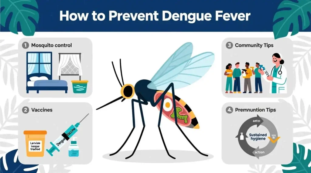
5.Prevention of Dengue Fever – Natural Remedies and Lifestyle Modifications
