

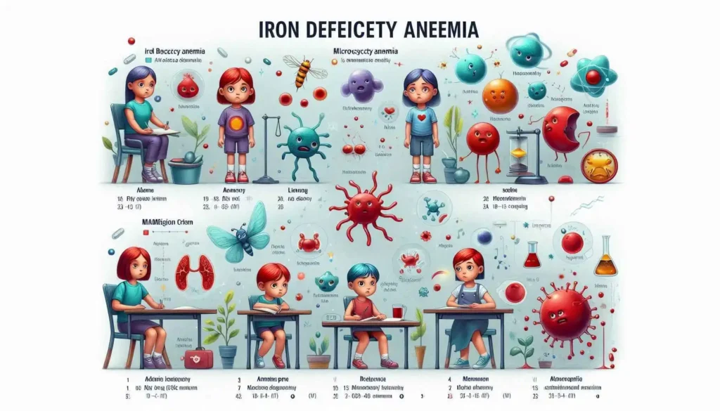
Microcytic Anemias: Causes, Symptoms, Diagnosis, and Treatments
“Anemia is not a disease; it is a clue that something in the body’s blood‑making machinery is out of balance.” — Dr. William A. Frank, MD, Hematology
Introduction
When we hear the word anemia, we often picture a patient who looks pale and feels constantly fatigued. Yet anemia is a broad umbrella that covers many distinct disorders, each with its own pathophysiology and therapeutic approach. Micro‑cytic anemia—characterized by red blood cells (RBCs) that are smaller than normal (mean corpuscular volume < 80 fL)—is one of the most common sub‑types. In this article we will explore, in depth, the major causes, typical symptoms, diagnostic work‑up, and evidence‑based treatments for micro‑cytic anemias. By the end, you should feel confident in recognizing and managing these conditions in a clinical setting.
What Is a Microcytic Anemia?

Microcytic anemia is a common type of anemia characterized by the presence of abnormally small red blood cells (erythrocytes) in the bloodstream. The term “microcytic” literally translates to “small cell,” referring to the diminished size of these oxygen-carrying cells. When red blood cells are smaller than normal, they may also contain less hemoglobin – the protein responsible for binding and transporting oxygen from the lungs to the body’s tissues. This reduced oxygen-carrying capacity can lead to various symptoms associated with anemia, such as fatigue, weakness, shortness of breath, and pallor.
Diagnostic Laboratory Hallmarks:
The diagnosis of microcytic anemia is primarily established through a complete blood count (CBC) test, which provides detailed information about red blood cell morphology. Microcytic anemia is defined by two key laboratory hallmarks:
| Parameter | Typical Finding | Explanation |
| Mean Corpuscular Volume (MCV) | < 80 fL (small red cells) | The MCV is the most crucial parameter, measuring the average size of individual red blood cells. A value less than 80 femtoliters (fL) definitively indicates that the red blood cells are microcytic, or smaller than their normal range. |
| Mean Corpuscular Hemoglobin Concentration (MCHC) | Often normal or slightly low | The MCHC measures the average concentration of hemoglobin within a red blood cell. In microcytic anemias, while the cells are small, the concentration of hemoglobin within them may still be relatively normal, or only mildly reduced. A significantly low MCHC would indicate hypochromia (pale red cells). |
Beyond the Definition: Uncovering the Root Cause
While a low MCV clearly identifies that the red cells are small, it does not reveal why they are small. Determining the underlying cause of microcytic anemia is a critical step, as the treatment and management strategies vary significantly depending on the etiology.
The formation of hemoglobin, and thus the proper size of red blood cells, relies on several key components, primarily iron and globins (protein chains). Issues with the production or appropriate utilization of these components often lead to microcytic red blood cells. The underlying causes of microcytic anemia fall into a handful of well-characterized categories, each with a distinct set of additional laboratory clues and clinical presentations that help differentiate them. Identifying these specific causes – such as iron deficiency, thalassemias, or anemia of chronic disease – is essential for effective patient care.
Understanding the underlying causes of anemia is paramount for accurate diagnosis and effective treatment. While anemia can stem from a myriad of factors, the conditions detailed below represent some of the major and most frequently encountered etiologies. This section provides a concise overview of their unique pathogenic mechanisms, followed by a comparative table (presented later) that highlights their key distinguishing features for clinical differentiation.
Major Causes of Anemia
1. Iron Deficiency Anemia (IDA)
- Pathogenesis: IDA typically arises when the body’s iron stores are insufficient to meet the demands for hemoglobin synthesis. This critical deficiency can be attributed to several primary mechanisms:
- Inadequate Iron Intake: Often due to poor dietary habits, vegetarian or vegan diets without proper supplementation, or limited access to iron-rich foods.
- Chronic Blood Loss: The most common cause in adults, leading to a continuous depletion of iron. Significant sources include gastrointestinal bleeding (e.g., ulcers, polyps, cancer, chronic NSAID use, parasitic infections like hookworm), heavy menstrual bleeding (menorrhagia), or frequent blood donations.
- Malabsorption: Conditions that impair the absorption of dietary iron, primarily in the duodenum. Examples include celiac disease, inflammatory bowel disease, Helicobacter pylori infection, gastrectomy, or bariatric surgery. The diminished iron availability directly compromises the production of functional hemoglobin, essential for oxygen transport.
2. Thalassemia Syndromes
- Pathogenesis: Thalassemia refers to a group of inherited blood disorders characterized by genetic mutations that impair or reduce the synthesis of one or more of the globin chains (alpha or beta) that constitute hemoglobin.
- These specific gene defects lead to an imbalanced production of hemoglobin, where the unaffected globin chains accumulate and precipitate within red blood cell precursors, causing cellular damage and premature destruction of red blood cells (ineffective erythropoiesis and hemolysis). The severity varies greatly depending on the specific mutation and the number of affected genes, ranging from asymptomatic carriers to severe, transfusion-dependent anemia.
3. Anemia of Chronic Disease (ACD) / Anemia of Inflammation
- Pathogenesis: ACD is a common anemia seen in individuals with chronic inflammatory conditions, infections, or malignancies. Its development is complex and primarily mediated by inflammatory cytokines (e.g., IL-6, TNF-alpha).
- These cytokines lead to an upregulation of hepcidin, a key regulator of iron metabolism. Hepcidin then causes the sequestration of iron in macrophages and hepatocytes, preventing its release into the circulation for erythropoiesis.
- Furthermore, cytokines can also decrease erythropoietin (EPO) production by the kidneys and render the bone marrow less responsive to EPO, further impairing red blood cell production. Essentially, iron is “locked away” from the developing red blood cells, even if total body iron stores are adequate or elevated.
4. Sideroblastic Anemia
- Pathogenesis: Sideroblastic anemia is characterized by defective heme synthesis within the mitochondria of erythroid precursors in the bone marrow. Heme is a crucial component of hemoglobin responsible for binding oxygen.
- Due to this defect, iron cannot be properly incorporated into the heme molecule and thus accumulates within the mitochondria, forming characteristic iron-laden ring sideroblasts (erythroid precursors with a collar of iron granules surrounding the nucleus).
- This ineffective utilization of iron leads to impaired hemoglobin production and ineffective erythropoiesis. Sideroblastic anemia can be inherited (congenital) or acquired, with acquired forms often associated with myelodysplastic syndromes, chronic alcohol abuse, certain drugs (e.g., isoniazid), or lead poisoning.
5. Lead Poisoning (Less Common but Serious)
- Pathogenesis: While less common today due to reduced environmental exposure, lead poisoning can cause anemia by directly interfering with several key enzymes in the heme synthesis pathway.
- Lead inhibits enzymes such as aminolevulinate dehydratase (ALA-D) and ferrochelatase. The inhibition of ferrochelatase specifically prevents the incorporation of iron into protoporphyrin to form heme.
- This disruption leads to the accumulation of heme precursors (e.g., protoporphyrin, ALA), which can be toxic, and ultimately results in reduced hemoglobin production. Lead can also directly damage red blood cell membranes, leading to premature destruction (hemolysis) and characteristic basophilic stippling visible on blood smears. Exposure typically occurs through old paint, contaminated water, or certain industrial settings.
Each of these etiologies presents with distinct clinical and laboratory features, emphasizing the critical role of comprehensive diagnostic evaluation in determining the specific cause of anemia and guiding appropriate therapeutic interventions. The subsequent comparative table will further delineate these differences to aid in accurate diagnosis.
| Condition | Primary Cause | Typical MCV (fL) | Iron Studies* | Ferritin (ng/mL) | Key Morphology | First‑Line Treatment |
| Iron‑Deficiency Anemia | Dietary deficiency / chronic blood loss | 70‑80 | Low serum iron, high TIBC | Low (<30) | Micro‑cytosis, hypochromia | Oral ferrous sulfate (or IV iron if intolerant) |
| β‑Thalassemia Minor | Heterozygous β‑globin gene mutation | 60‑75 | Normal/low serum iron, normal TIBC | Normal or high | Target cells, basophilic stippling | Usually none; folic acid if needed |
| Anemia of Chronic Disease | Inflammatory cytokines (e.g., rheumatoid arthritis) | 70‑80 | Low serum iron, low TIBC | Normal or high | Normo‑ or micro‑cytic, sometimes anisocytosis | Treat underlying disease; consider erythropoiesis‑stimulating agents |
| Sideroblastic Anemia | Defective δ‑ALA synthase, alcohol, drugs, hereditary | 70‑80 | Normal/high serum iron, low TIBC | High | Ring sideroblasts in marrow | Pyridoxine (B6), remove offending agents |
| Lead Poisoning | Occupational/ environmental exposure | 60‑80 | Low serum iron, low TIBC | Normal/high | Basophilic stippling | Chelation therapy (EDTA, dimercaprol) |
*Iron studies: serum iron, total iron‑binding capacity (TIBC), transferrin saturation, and ferritin.
Clinical Presentation: Symptoms to Watch For in Microcytic Anemias
Understanding the clinical presentation is paramount in the diagnostic workup of microcytic anemias. While these conditions, characterized by smaller-than-normal red blood cells, share many of the classic anemia signs reflecting the body’s struggle with reduced oxygen delivery to tissues, certain distinguishing features and historical clues can significantly narrow the differential diagnosis and guide further investigation.
Shared General Symptoms of Anemia:
While non-specific, the following symptoms are universally present in varying degrees across all microcytic anemias, indicating the underlying hypoxia:
- Fatigue, Weakness: These are the most common and often the earliest complaints. Patients report persistent tiredness, lack of energy, and reduced stamina, even with minimal exertion. This results directly from insufficient oxygen supply to muscles and organs.
- Dyspnea on exertion: Shortness of breath, particularly during physical activity, occurs as the body attempts to compensate for reduced oxygen-carrying capacity by increasing respiratory rate. In severe cases, dyspnea may be present even at rest.
- Pallor (conjunctival, mucosal): A noticeable paleness of the skin, mucous membranes (especially inside the mouth), and the conjunctiva of the eyes is a tell-tale sign of anemia. This is due to reduced oxyhemoglobin in the blood vessels near the surface.
Distinguishing Features by Anemia Type:
Beyond these general symptoms, specific findings during history taking and physical examination can provide crucial insights:
| Symptom/Sign | Typical in Iron Deficiency Anemia (IDA) | Typical in Thalassemia | Typical in Anemia of Chronic Disease (ACD) | Typical in Sideroblastic Anemia |
| Fatigue, Weakness | ✓ | ✓ | ✓ | ✓ |
| Dyspnea on exertion | ✓ | ✓ | ✓ | ✓ |
| Pallor (conjunctival, mucosal) | ✓ | ✓ | ✓ | ✓ |
| Pica (craving ice) | Common: The compulsive craving and consumption of non-nutritive substances, particularly ice (pagophagia), is a highly specific, though not fully understood, symptom of IDA. It’s less commonly seen with other forms of pica like geophagia (clay) or amylophagia (starch). | Rare | Rare | Rare |
| Koilonychia (spoon nails) | Common: Characterized by soft, brittle nails that flatten and then develop a concave, spoon-like depression. This is a classic, though late, sign of chronic iron deficiency, reflecting impaired keratin synthesis. | Rare | Rare | Rare |
| Splenomegaly | Rare (unless very severe or due to comorbid conditions like portal hypertension) | Frequent (especially β-thalassemia): Occurs due to increased extramedullary hematopoiesis (blood cell production outside the bone marrow) and increased destruction of abnormal red blood cells by the spleen. Can range from mild to massive. | Rare | Possible (if severe, particularly in myelodysplastic syndrome-associated types or lead poisoning) |
| Bone pain / facial bone deformities | Rare | Typical (especially β-thalassemia major): Severe forms, like thalassemia major, lead to marked ineffective erythropoiesis. The body’s attempt to produce red blood cells causes massive expansion of the bone marrow. This can result in thinning of cortical bones, widening of diploic spaces, and characteristic facial changes (e.g., “chipmunk facies” with prominent frontal bones and maxilla), as well as chronic bone pain. | Rare | Rare |
| Neurologic symptoms (peripheral neuropathy) | Rare (though restless legs syndrome is frequent) | Rare | Rare | If due to lead: Lead poisoning, a cause of acquired sideroblastic anemia, often presents with peripheral neuropathy (e.g., “lead palsy” affecting motor nerves), abdominal pain, and cognitive issues. |
| Gastrointestinal symptoms (melena, hemorrhoids) | Suggests chronic blood loss: These are not direct symptoms of IDA itself, but rather common underlying causes of iron deficiency. A thorough history of potential blood loss (e.g., heavy menstrual periods, gastrointestinal bleeding, frequent nosebleeds) is crucial. | Rare | Rare | Rare |
Clinical Application and Differential Diagnosis:
When obtaining a patient’s history, these nuanced clues are invaluable for narrowing the differential considerably. For instance:
- A young adult with a family history of anemia, mild microcytosis, and splenomegaly strongly suggests a genetic hemolytic disorder like thalassemia trait rather than simple iron deficiency. The family history points to an inherited condition, and splenomegaly is a hallmark of increased red cell destruction or extramedullary hematopoiesis seen in thalassemia.
- Conversely, a premenopausal woman with heavy menstrual bleeding, unexplained fatigue, and a craving for ice points significantly towards iron deficiency anemia.
- A patient with long-standing rheumatoid arthritis presenting with mild to moderate anemia, despite adequate iron stores, would primarily suggest anemia of chronic disease (ACD).
- An individual with occupational exposure to lead experiencing abdominal pain and neurological symptoms alongside microcytic anemia should raise strong suspicion for sideroblastic anemia secondary to lead poisoning.
In essence, while the initial symptoms of microcytic anemia may overlap, a detailed medical history, including family history, dietary habits, occupational exposures, and a careful physical examination for specific signs, provides critical direction for further diagnostic testing, such as iron studies, hemoglobin electrophoresis, and bone marrow biopsy, to definitively identify the underlying cause.
Comprehensive Diagnostic Workup for Microcytic Anemia
A systematic, step-wise approach is paramount in the evaluation of microcytic anemia. This structured methodology not only helps to avoid unnecessary investigations and reduce patient burden but also ensures accurate identification of the underlying etiology, which is crucial for initiating appropriate and effective treatment. Microcytic anemia is characterized by red blood cells that are smaller than normal (low Mean Corpuscular Volume, MCV) and is most commonly caused by iron deficiency, thalassemia, or anemia of chronic disease.
