

What Causes Lung Cancer? Symptoms, Diagnosis & Latest Treatment Advances
“Cancer is a word, not a sentence.” – John Diamond
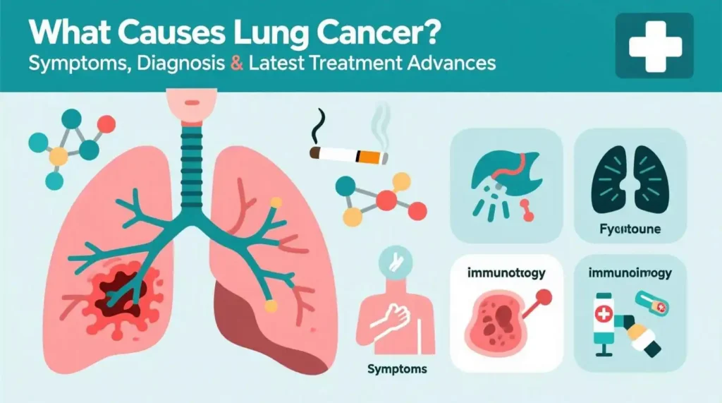
We know that lung cancer remains one of the most formidable health challenges of our time. In this article we will walk through the disease’s major causes, symptoms, diagnostic pathways, and treatment options, providing a clear, evidence‑based picture for patients, families, and health‑care professionals alike. Throughout, we will use the first‑person plural (“we”) to emphasize that tackling lung cancer is a collective effort—one that requires the cooperation of researchers, physicians, public‑health officials, and the broader community.
1. Why Does Lung Cancer Develop?
Lung cancer arises when normal lung cells acquire genetic mutations that disrupt the tightly regulated balance between cell growth and death. These mutations can be sporadic (arising spontaneously) or induced by external carcinogens. Below is a concise summary of the most prominent risk factors.

| Risk Factor | Relative Risk Increase | Mechanism |
| Tobacco smoking (current) | 20–30× higher vs. never‑smoker | Direct DNA adduct formation, chronic inflammation |
| Former smoker (≤15 pack‑years) | 5–10× higher | Residual DNA damage; risk declines after cessation |
| Radon gas exposure | 2–3× higher | Alpha particle radiation causing DNA double‑strand breaks |
| Asbestos & occupational pollutants | 1.5–2× higher | Fibers induce chronic inflammation & oxidative stress |
| Air pollution (PM₂.₅) | 1.2–1.5× higher | Particulate‑induced oxidative DNA damage |
| Genetic predisposition (e.g., EGFR, KRAS mutations) | Variable | Inherited mutations predispose cells to malignant transformation |
| Previous thoracic radiation therapy | 1.5–2× higher | Radiation‑induced DNA lesions |
Key take‑away: While smoking accounts for ~85 % of all cases, a substantial minority of lung cancers arise in never‑smokers—particularly women, younger individuals, and people with significant radon exposure or genetic susceptibility.
2. Recognizing the Signs: Common Symptoms
Early lung cancer is often silent. By the time symptoms appear, the disease may already be locally advanced or metastatic. Below we list the most frequently encountered clinical presentations, grouped by organ system.
2.1 Respiratory‑related symptoms
- Persistent cough (new or worsening, especially if “smoker’s cough” changes character)
- Hemoptysis (coughing up blood or rust‑colored sputum)
- Dyspnea (shortness of breath at rest or on exertion)
- Wheezing or noisy breathing
One of the most insidious aspects of lung cancer is its often-silent nature in its early stages. Unlike many other diseases that present with clear warning signs from their inception, early lung cancer is frequently asymptomatic and remains undetected. This is primarily because the lungs themselves have few nerve endings, meaning a small, localized tumor may not cause pain or discomfort.
Consequently, by the time noticeable symptoms appear, the disease may already be locally advanced, meaning it has spread within the lung or to nearby structures, or it may even be metastatic, having spread to distant parts of the body. This late presentation significantly impacts the prognosis and the complexity of treatment. It’s crucial for individuals, especially those at high risk, to be aware of the subtle yet significant changes that could signal the presence of lung cancer.
Below we detail the most frequently encountered clinical presentations, grouped by the organ system primarily affected, beginning with those directly related to the respiratory system.
As lung cancer originates in the lungs, the most common and often the first symptoms to emerge are those affecting the respiratory system. These symptoms are caused by the tumor’s growth within the airways, its compression of nearby structures, or its invasion into lung tissue. It’s important to note that while these symptoms can also be indicative of less serious conditions like infections or chronic obstructive pulmonary disease (COPD), their persistence, worsening, or unusual character necessitates medical evaluation.
Persistent Cough (new or worsening, especially if “smoker’s cough” changes character): This is the most common symptom of lung cancer. It refers to a cough that does not go away after a few weeks, or one that progressively worsens over time. For individuals with a pre-existing “smoker’s cough,” a significant warning sign is a change in the character of this chronic cough. This might involve becoming more frequent, deeper, harsher, more painful, or producing different amounts or types of mucus. The cough can be dry or produce sputum, and it’s often the result of the tumor irritating the airways, causing inflammation, or obstructing airflow.
Hemoptysis (coughing up blood or rust-colored sputum): This is an alarming symptom that should always prompt immediate medical attention, regardless of the amount of blood. Hemoptysis can manifest as streaks of bright red blood mixed with phlegm, rusty-colored sputum (indicating older blood), or, less commonly, larger amounts of blood. This occurs when the tumor invades or erodes into blood vessels within the lung or airways, causing them to bleed. Even small amounts of blood are significant and should not be dismissed.
Dyspnea (shortness of breath at rest or on exertion): This refers to difficulty breathing or a feeling of not getting enough air. It often has a progressive onset, meaning it starts subtly and gradually worsens over time. Initially, it might only be noticeable during physical exertion (like climbing stairs or walking long distances), but as the disease progresses, it can occur with minimal activity or even at rest. Shortness of breath can be caused by the tumor blocking a major airway, fluid accumulation around the lungs (pleural effusion), the collapse of part of the lung (atelectasis) due to obstruction, or the tumor pressing on nerves that control breathing.
Wheezing or Noisy Breathing: Wheezing is a high-pitched, whistling sound produced during breathing, particularly when exhaling. It occurs when airways become narrowed or blocked, forcing air through a constricted passage. In lung cancer, this can happen if a tumor is growing within an airway or pressing on an airway from the outside. While wheezing is common in conditions like asthma or COPD, new-onset wheezing, especially if it’s localized to one side of the chest (unilateral wheezing) and persistent, can be a sign of a cancerous obstruction.
2.2 Systemic symptoms
- Unexplained weight loss (≥5 % of body weight over 6–12 months)
- Anorexia (loss of appetite)
- Fatigue or generalized weakness
- Bone pain (particularly in the spine, ribs, or pelvis)
Systemic symptoms refer to the manifestations of a disease or condition that affect the entire body, rather than being confined to a specific organ or system. In the case of certain medical conditions, such as cancer or infections, these symptoms may be indicative of a more severe underlying issue. The following are some of the common systemic symptoms that may arise:
Unexplained weight loss: A loss of 5% or more of one’s body weight over a period of 6 to 12 months, without any specific changes in diet or exercise habits, may be a cause for concern. This weight loss can be a result of various factors, including the body’s inability to absorb nutrients or the increased metabolic demands of an underlying disease.
Anorexia: Anorexia, or loss of appetite, can be another sign of a systemic issue. This may manifest as a general disinterest in food or a reduced ability to enjoy the taste of food. Anorexia can lead to malnutrition and further exacerbate the underlying condition.
Fatigue or generalized weakness: A persistent feeling of tiredness or lack of energy, even after rest, can be a symptom of various systemic conditions. This may be due to the body’s increased energy demands or the depletion of essential nutrients.
Bone pain: Pain in the bones, particularly in the spine, ribs, or pelvis, can be indicative of a more severe underlying condition. This pain may be due to the spread of cancer cells to the bones, or an infection that has affected the bone marrow.
Systemic symptoms should not be ignored, as they may be indicative of a more severe underlying condition. If you experience any of these symptoms, it is essential to consult with a healthcare professional for a proper evaluation and diagnosis.
2.3 Paraneoplastic phenomena
Lung tumors, especially small‑cell carcinoma, can secrete hormones or cytokines that cause distant effects:
- Hypercalcemia (due to parathyroid hormone‑related peptide)
- Hypertrophic pulmonary osteoarthropathy (clubbing, periostitis)
- Ectopic ACTH production (Cushing’s syndrome)
Clinical tip: Because many of these signs overlap with common respiratory infections, we advise clinicians to maintain a low threshold for imaging in patients over 40 with a smoking history or any persistent respiratory complaint.
Lung tumors, particularly small cell carcinoma, have the ability to secrete hormones or cytokines that can cause distant effects in the body. These paraneoplastic phenomena can lead to a variety of symptoms and complications that may not be directly related to the primary tumor or its metastasis. Some of the common paraneoplastic effects associated with lung tumors include:
Hypercalcemia: This condition occurs when the cancer cells produce a protein called parathyroid hormone-related peptide (PTHrP). PTHrP mimics the action of parathyroid hormone, leading to an increase in calcium levels in the blood. This can cause symptoms such as fatigue, weakness, confusion, and constipation. In severe cases, hypercalcemia can lead to kidney stones, bone pain, and even coma.
Hypertrophic Pulmonary Osteoarthropathy (HPOA): Also known as “clubbing,” HPOA is a condition in which the fingers and toes become swollen and misshapen. This is due to the overgrowth of connective tissue and bone in the extremities. HPOA can also cause pain and tenderness in the joints, particularly those of the hands and feet. The exact mechanism behind HPOA is not well understood, but it is thought to be related to the production of cytokines by the cancer cells.
Ectopic ACTH Production: In some cases, lung tumors can produce a hormone called adrenocorticotropic hormone (ACTH) outside of the pituitary gland. This is known as ectopic ACTH production. ACTH stimulates the adrenal glands to produce cortisol, a stress hormone. An excess of cortisol can lead to Cushing’s syndrome, a condition characterized by weight gain, muscle weakness, easy bruising, and other symptoms.
Clinical Tip:
Due to the overlapping nature of paraneoplastic symptoms with common respiratory infections, it is essential for clinicians to maintain a low threshold for imaging in patients over 40 with a smoking history or any persistent respiratory complaints. Early detection and diagnosis of lung tumors can significantly improve the prognosis and treatment outcomes for affected individuals. Imaging studies, such as chest X-rays or CT scans, can help identify the presence of lung tumors and guide further diagnostic and treatment strategies.
3. How Do We Diagnose Lung Cancer?
A systematic, stepwise approach ensures accurate staging and guides therapeutic decisions. Below is the typical diagnostic algorithm we follow.
3.1 Initial Assessment
- History & Physical Examination – Document smoking exposure, occupational hazards, family history, and symptom chronology.
- Chest Radiograph – Often the first imaging study; may reveal a solitary pulmonary nodule, mass, or hilar enlargement.
How Do We Diagnose Lung Cancer?
A systematic, stepwise approach ensures accurate staging and guides therapeutic decisions. Below is the typical diagnostic algorithm we follow:
In the initial assessment of a patient suspected of having lung cancer, it is crucial to gather a detailed history and perform a thorough physical examination. This includes documenting smoking exposure, occupational hazards, family history, and the chronology of symptoms. This information will help guide further diagnostic testing and treatment planning.
Chest Radiograph:
The chest radiograph is often the first imaging study performed when lung cancer is suspected. It may reveal various findings indicative of lung cancer, such as a solitary pulmonary nodule, a mass, or hilar enlargement. A solitary pulmonary nodule is a small, round, or oval-shaped opacity measuring up to 3 cm in diameter. A mass is a larger opacity, usually greater than 3 cm in diameter. Hilar enlargement refers to an increase in the size of the lymph nodes in the hilum of the lung, which may indicate the presence of lung cancer or another disease process.
If a suspicious finding is identified on the chest radiograph, further imaging studies may be ordered to better characterize the lesion, such as a computed tomography (CT) scan or positron emission tomography (PET) scan.
Computed Tomography (CT) Scan:
A CT scan is a non-invasive imaging study that uses X-rays and computer technology to create detailed images of the internal structures of the body. It can provide more detailed information about the size, shape, and location of a lung mass or nodule, as well as any associated lymph node enlargement. CT scans can also help identify the presence of metastatic disease in other organs.
Positron Emission Tomography (PET) Scan:
A PET scan is a nuclear medicine imaging technique that can help detect cancerous cells based on their increased metabolic activity. It involves the injection of a small amount of a radioactive tracer, which is taken up by cells with high metabolic activity, such as cancer cells. The PET scan can help identify the primary tumor, as well as any metastatic disease in other organs.
Tissue Sampling:
Once a suspicious lesion has been identified on imaging studies, tissue sampling is necessary to confirm the diagnosis of lung cancer. This can be achieved through various methods, including:
Bronchoscopy: A procedure in which a thin, flexible tube is inserted through the mouth or nose and into the airways to obtain a tissue sample from the lung.
Transthoracic needle aspiration (TTNA): A procedure in which a needle is inserted through the chest wall to obtain a tissue sample from the lung.
Surgical biopsy: A procedure in which a surgeon removes a portion of the lung tissue for examination under a microscope.
Staging:
After a diagnosis of lung cancer has been confirmed, staging is performed to determine the extent of the disease. This involves assessing the size of the tumor, the involvement of lymph nodes, and the presence of distant metastases. Staging is crucial for guiding treatment decisions and estimating the prognosis.
The diagnosis of lung cancer involves a systematic, stepwise approach that includes initial assessment, imaging studies, tissue sampling, and staging. This comprehensive evaluation ensures accurate staging and guides therapeutic decisions to provide the best possible care for patients with lung cancer.
3.2 Advanced Imaging
| Modality | Indications | What It Shows |
| Low‑dose CT (LDCT) | Lung cancer screening (high‑risk individuals) | Detects nodules as small as 4 mm |
| Contrast‑enhanced CT | Staging, surgical planning | Tumor size, mediastinal nodes, invasion of chest wall/diaphragm |
| PET‑CT (18F‑FDG) | Metabolic assessment, detection of occult metastases | Hypermetabolic lesions; helps differentiate benign from malignant nodules |
| MRI brain | Suspected or confirmed stage III‑IV disease | Detects brain metastases, especially in adenocarcinoma |
3.3 Tissue Diagnosis
Obtaining a histologic specimen is mandatory before definitive therapy. Options include:
- Bronchoscopy with endobronchial biopsy (for centrally located lesions)
- CT‑guided percutaneous needle biopsy (peripheral nodules)
- Navigational bronchoscopy (advanced technique for small peripheral lesions)
- Surgical biopsy (via video‑assisted thoracoscopic surgery, VATS) when minimally invasive methods fail
During sampling, we also request molecular profiling (EGFR, ALK, ROS1, BRAF, KRAS, PD‑L1, etc.) to identify actionable targets.
Tissue diagnosis is a crucial step in the management of lung cancer, as it provides valuable information for determining the most appropriate treatment strategy. Obtaining a histologic specimen is mandatory before initiating definitive therapy. There are several methods available to obtain tissue samples from the tumor, depending on the location and characteristics of the lesion.
Bronchoscopy with endobronchial biopsy: This method is suitable for centrally located lesions, which are easily accessible through the bronchial tree. A bronchoscope is inserted through the patient’s nose or mouth and guided to the target area, where a small piece of tissue is collected using forceps or other instruments.
CT guided percutaneous needle biopsy: This technique is used for peripheral nodules, which are located further from the central airways. A needle is guided through the patient’s chest wall under CT guidance to reach the target lesion, and a small piece of tissue is obtained for histological analysis.
Navigational bronchoscopy: This is an advanced technique used for small peripheral lesions that are difficult to reach with standard bronchoscopy. The procedure involves the use of a virtual bronchoscopy system, which creates a three-dimensional map of the patient’s airways based on CT images. A bronchoscope is then navigated through the airways using the virtual map to reach the target lesion, and a biopsy is performed.
Surgical biopsy via video-assisted thoracoscopic surgery (VATS): If minimally invasive methods fail to obtain a sufficient tissue sample, a surgical biopsy may be performed. This involves making small incisions in the chest wall and inserting a thoracoscope to visualize the lung and obtain a tissue sample.
During the sampling process, we also request molecular profiling of the tissue specimen to identify actionable targets. Molecular profiling involves the analysis of specific genes and proteins that are commonly altered in lung cancer, such as EGFR, ALK, ROS1, BRAF, KRAS, and PD-L1. This information can help guide targeted therapies and immunotherapies, which have shown promising results in the treatment of lung cancer.
Tissue diagnosis is an essential step in the management of lung cancer, and various methods are available to obtain histologic specimens. Molecular profiling is also performed to identify actionable targets and guide personalized treatment strategies.
